


1. What Is PD-1?
PD-1 (Programmed Death-1) is a protein expressed on the surface of T cells. When tumor cells highly express PD-L1, PD-L1 binds to PD-1 and transmits a “stop killing” signal to T cells, allowing tumors to evade immune attack. PD-1-targeted drugs are primarily inhibitors (anti-PD-1 antibodies) that block the binding of PD-1 to PD-L1, restoring the ability of T cells to recognize and kill tumor cells.
PD-1-targeted drug mechanism of action (figure caption)
There are two major directions in the iterative development of PD-1 for cancer immunotherapy: bispecific antibodies (such as PD-1/VEGF bispecific antibodies) and combination with ADC, chemotherapy, and anti-angiogenic agents.
2. What Is VEGFA?
VEGFA (Vascular Endothelial Growth Factor A) is secreted by tumor cells and other cells in the tumor microenvironment. Upon binding to VEGFR on vascular endothelial cells, it promotes endothelial cell proliferation and the formation of new blood vessels. Once solid tumors reach a certain size, they depend on neovascularization to obtain oxygen and nutrients, and use it to invade and metastasize.
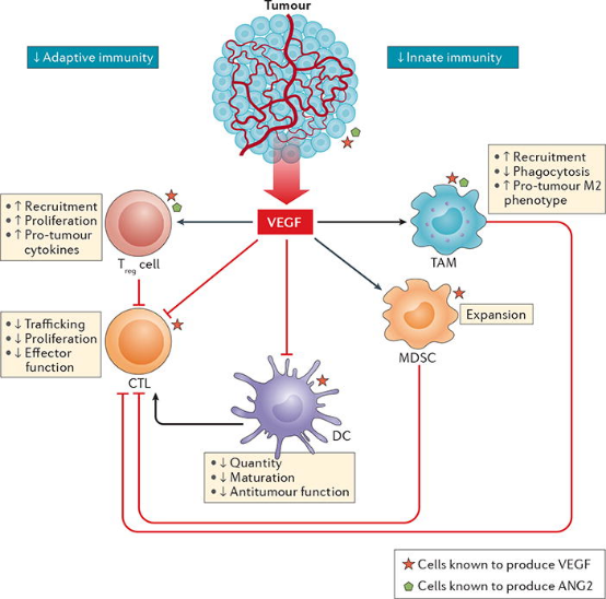
VEGFA-targeted drug mechanism of action: Anti-VEGF-A agents (such as bevacizumab) bind to VEGF-A, preventing it from activating its receptor, thereby inhibiting neovascularization and inducing regression of some existing tumor vasculature. In addition, vascular “normalization” may improve drug delivery and T cell infiltration—this is the mechanistic basis for combining “anti-angiogenic + immunotherapy” regimens.
3. What Is TROP2?
TROP2 (Trophoblast Cell-Surface Antigen 2), encoded by the TACSTD2 gene, is a type I transmembrane glycoprotein involved in processes such as calcium signaling. It is characterized by high expression in a variety of epithelial-derived tumors—including triple-negative breast cancer (TNBC), non-small cell lung cancer, urothelial carcinoma, and gastric cancer—with low expression in normal tissues, and high expression is often associated with poorer prognosis.
TROP2-targeted drug mechanism of action: The mainstream therapeutic format for TROP2 is ADC (antibody-drug conjugate): the antibody moiety binds to TROP2 on the tumor cell surface → internalization into the cell → cleavage of the linker inside the cell → release of topoisomerase I inhibitor payloads → killing of tumor cells. Different products differ in antibody, linker, payload, and DAR (drug-to-antibody ratio), resulting in markedly different efficacy and toxicity profiles.
The most common failure of humanized models is not “failure to knock in,” but insertion without expression: randomly inserted transgenes rely on exogenous strong promoters to drive expression, losing tissue specificity; in situ replacement often lands in transcriptionally silent regions. MingCeler’s EnhancerPlus analysis platform predicts regulatory element positions based on proprietary data and, combined with codon optimization, redesigns editing strategies to achieve near-endogenous expression levels. Through the TurboMice™ technology platform, multi-target homozygous humanized mice can be rapidly generated. We welcome all researchers to consult us.
References and Sources
- Ishida Y, Agata Y, Shibahara K, Honjo T. Induced expression of PD-1, a novel member of the immunoglobulin gene superfamily, upon programmed cell death. J Immunol. 1992;149(12):3881-3885.
- Yokosuka T, Takamatsu M, Kobayashi-Imanishi W, Hashimoto-Tane A, Azuma M, Saito T. Programmed cell death 1 forms negative costimulatory microclusters that directly inhibit T-cell receptor signaling by recruiting phosphatase SHP2. Nat Immunol. 2012;13(2):162-172.
- Hurwitz H, Fehrenbacher L, Novotny W, et al. Bevacizumab plus irinotecan, fluorouracil, and leucovorin for metastatic colorectal cancer. N Engl J Med. 2004;350(23):2335-2342.
- Fukumura D, Kloepper J, Amoozgar Z, Duda DG, Jain RK. Enhancing cancer immunotherapy using antiangiogenics: opportunities and challenges. Nat Rev Clin Oncol. 2018;15(5):325-340.
- Trerotola M, Relli V, Simeone P, Alberti S. TROP2: a tumor-associated calcium signal transducer. Int J Cancer. 2014;134(11):2547-2554.