


Gene editing alters genomic DNA sequence, but RNA splicing ultimately dictates which mature transcripts are produced from edited alleles, and whether those transcripts can generate functional proteins. The magnitude of loss-of-function in any gene-edited mouse model depends not on the presence of a DNA indel alone, but on final functional protein abundance.
A common and underrecognized problem: many CRISPR-edited mice show no observable phenotype despite confirmed Cas9 activity at the DNA level. In most cases, the root cause is not failed gene editing, but unanticipated disrupted or compensatory RNA splicing that preserves functional protein expression.
Fundamentals of RNA Splicing
Most protein-coding genes in the mouse genome are split genes: coding exons are separated by non-coding introns. After transcription, the primary transcript (pre-mRNA) undergoes splicing to remove introns and ligate exons into mature, translation-competent mRNA.
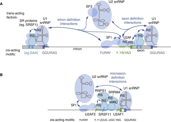
The spliceosome is assembled from U1, U2, U4/U6, U5 snRNPs and auxiliary proteins. It recognizes these core sequence elements:
| Element | Location and Conserved Features |
|---|---|
| 5′ splice site (donor site) | 5′ end of the intron, marked by the conserved dinucleotide GU |
| Branch point A | Inside the intron, roughly 20–50 nt upstream of the 3′ end |
| 3′ splice site (acceptor site) | 3′ end of the intron, conserved dinucleotide AG, preceded by a polypyrimidine tract |
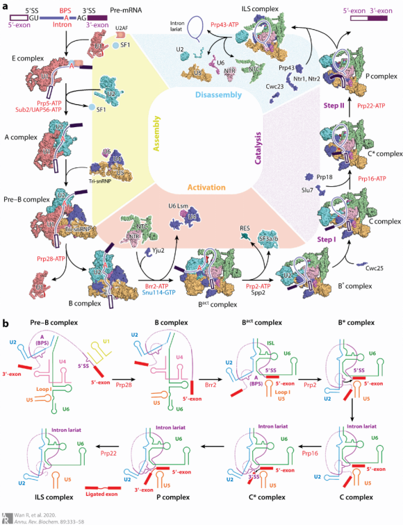
Following two transesterification reactions, the intron is released as a lariat structure and degraded, while adjacent exons are joined together.
Alternative Splicing
A single pre-mRNA can yield multiple transcript isoforms via distinct splicing patterns:
- Exon skipping: The exon is excluded, generating a protein lacking the corresponding domain.
- Intron retention: Frequently introduces a premature termination codon (PTC); such transcripts are commonly degraded via nonsense-mediated mRNA decay (NMD).
- Splice-site shift: Alters exon length, potentially triggering frameshifts or amino acid sequence changes.
- Mutually exclusive exons: Different isoforms retain separate exons, which may produce proteins with divergent functions.
For this reason, you must review all major transcript isoforms of your target gene before designing editing strategies, rather than only referencing the longest canonical RefSeq isoform.
How Splicing Shapes Gene-Edited Mouse Models
1. Reading Frame after Exon Deletion
Whole-body knockout often relies on deleting one or more exons. If the deleted region spans a multiple of three nucleotides and the remaining exons splice normally, the resulting truncated protein may retain partial function.
Design guidelines:
- Select key exons that cover a large fraction of the coding region.
- Verify that deletion induces a frameshift with an early downstream stop codon.
- Inspect every transcript isoform individually; do not validate only one isoform.
2. Targeting Splice Sites to Downregulate Protein Levels
Place sgRNA at the 5′ donor site of an exon–intron junction. Indels at this position disrupt the conserved GU motif, triggering:
- Failure of canonical splicing
- Recruitment of cryptic splice sites, producing transcripts with deletions or insertions
- Most abnormal transcripts carry PTCs and are cleared by NMD
- Reduced steady-state protein expression
This strategy creates an intermediate loss-of-function model between heterozygous deletion and full knockout. Important note: splice-site-edited alleles are not guaranteed to be null alleles. Functional validation using RT-PCR and western blot is mandatory.
3. Splicing Considerations for Conditional Knockout (cKO)
Standard flox designs place LoxP sites within introns flanking a critical exon. Cre-mediated excision of the exon creates a frameshift and gene inactivation.
LoxP insertion must satisfy all of these criteria:
- Do not disrupt the 5′ donor, branch point, polypyrimidine tract, or 3′ acceptor site
- Do not interfere with intronic regulatory elements or exonic splicing enhancers (ESEs)
- Avoid exon skipping or reduced expression before Cre recombination
If homozygous flox mice show complete loss of protein without Cre induction, first investigate splicing defects introduced by LoxP insertion, instead of attributing the phenotype directly to Cre leakage.
4. Artificial Intron Conditional Alleles
This design inserts a self-splicing artificial intron into the coding sequence:
| Status | Outcome |
|---|---|
| Pre-recombination | The artificial intron is precisely excised; the downstream coding sequence maintains the original reading frame and protein is expressed normally |
| Post-Cre recombination | The branch point and polypyrimidine region are removed, splicing fails; residual artificial intron sequence remains in mRNA, introducing PTC and frameshift, and transcripts are degraded by NMD |
Systems such as DECAI / AIv4 support one-step conditional allele generation; SCON is a shorter equivalent cassette that can be knocked into zygotes in a single step using ssODN donors.
When selecting these systems, evaluate two key metrics: precise splicing efficiency in the unrecombined state, and mRNA clearance efficiency after recombination. The former ensures clean baseline phenotypes; the latter determines knockout completeness.
5. Splicing Alterations from Small Edits
Even minor modifications can reshape splicing behaviour:
- Base edits or point mutations disrupting canonical GU/AG splice signals
- Synonymous mutations that alter ESEs or activate cryptic splice sites
- Small insertions that make exon length non-triple or introduce new donor/acceptor dinucleotides
- Homology donor arms or LoxP sequences that contain recognizable splice signals
Synonymous mutations therefore cannot be assumed to be functionally silent. Donor templates should undergo splice-site scanning before synthesis.
Closing Takeaway
RNA splicing describes the process by which the spliceosome removes introns and ligates exons from pre-mRNA in eukaryotic cells. Alternative splicing enables a limited set of genes to generate diverse protein isoforms, serving as a core layer of gene expression regulation.
Splicing is a critical variable to control for constitutive KO, cKO and KI mouse design. It can also be harnessed intentionally: splice-site targeting for protein knockdown, artificial introns for conditional alleles, and exon-skipping reporter mice to quantify splicing modulation. The success of your editing design ultimately depends on the final transcript profile and protein expression level.
References
- Wilkinson ME, Charenton C, Nagai K. How is precursor messenger RNA spliced by the spliceosome? Annual Review of Biochemistry. 2020;89:359–388. doi:10.1146/annurev-biochem-013118-111024
- Rogalska ME, Vivori C, Valcárcel J. Regulation of pre-mRNA splicing: roles in physiology and disease, and therapeutic prospects. Nature Reviews Genetics. 2023;24:251–269. doi:10.1038/s41576-022-00556-8
- Ule J, Blencowe BJ. Alternative splicing regulatory networks: functions, mechanisms, and evolution. Molecular Cell. 2019;76:329–345. doi:10.1016/j.molcel.2019.09.017
- Shenasa H, Bentley DL. Pre-mRNA splicing and its cotranscriptional connections. Trends in Genetics. 2023;39:672–685. doi:10.1016/j.tig.2023.04.008