




What Is Niemann-Pick Disease?
Niemann-Pick Disease (NPD) is an autosomal recessive lysosomal storage disorder, also referred to as sphingomyelin-cholesterol lipidosis. Based on pathogenic mechanisms and clinical manifestations, NPD is divided into two major subgroups: Type A/B and Type C.
Type A/B (Acid Sphingomyelinase Deficiency)
- Type A (acute neurological form): Onset occurs at 3–6 months after birth, characterized by hepatosplenomegaly, feeding difficulties, severe cachexia, and progressive neurodevelopmental regression. Approximately half of patients present a cherry-red spot on the fundus; most patients pass away before 3–4 years of age.
- Type B (non-neurological/visceral form): Symptoms manifest later, dominated by hepatosplenomegaly with intact intellectual function and absence of neurological lesions. Patients can survive into adulthood but frequently develop complications including interstitial lung disease and thrombocytopenia.
Type C (Cholesterol Transport Disorder)
Onset ages vary widely, ranging from perinatal period to adulthood. Hallmark neurological manifestations observed in children and adolescents include vertical supranuclear gaze palsy, ataxia, dysarthria, cataplexy with laughter, and cognitive impairment.
Epidemiological data show the incidence of NPD-A/B ranges from 1 in 250,000 to 1 in 44,960 worldwide, while NPC incidence is 1 in 150,000 to 1 in 120,000. Asian populations have a lower overall prevalence, with Type A accounting for roughly 85% of all NPD cases.
Pathogenesis
Niemann-Pick Type A/B (NPD-A/B)
NPD-A/B arises from loss-of-function variants in the SMPD1 gene mapped to chromosomal locus 11p15.1-p15.4, which encodes acid sphingomyelinase (ASM). ASM resides within lysosomes and catalyzes the hydrolysis of sphingomyelin into ceramide and phosphocholine. Pathogenic mutations drastically reduce ASM activity: <5% residual activity in Type A patients and 5%–10% in Type B patients, blocking normal sphingomyelin breakdown.
Deficient ASM activity triggers massive sphingomyelin accumulation in the mononuclear phagocyte system and central nervous system, generating characteristic foam cells (Niemann-Pick cells). As a core structural component of cell and organelle membranes, accumulated sphingomyelin causes multi-organ damage: hepatosplenomegaly in liver and spleen, interstitial pulmonary lesions, and neuronal dysfunction with progressive neurodegeneration in the brain. Additionally, insufficient ceramide production disrupts cellular signaling and apoptotic regulation, further accelerating disease progression.
Niemann-Pick Type C (NPC)
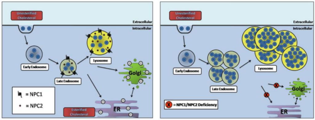
NPC is caused by mutations in either NPC1 or NPC2, which disrupt lysosomal cholesterol trafficking. Under physiological conditions, NPC2 delivers intralysosomal cholesterol to the membrane-bound NPC1 protein for efflux to other cellular compartments. Loss of NPC1/NPC2 function traps cholesterol and sphingolipids inside lysosomes while depleting cholesterol stores in the endoplasmic reticulum and other organelles.
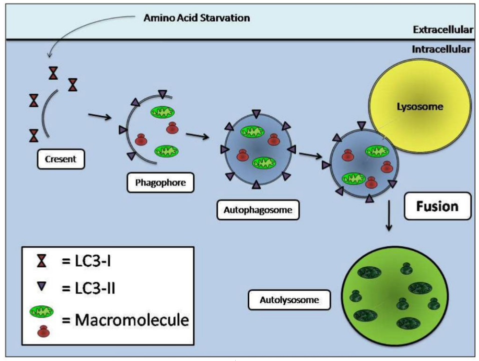
Impaired lipid transport aberrantly activates autophagic signaling. Accumulated lysosomal sphingolipids (e.g., ceramide) stimulate the Class III PI3K/Beclin-1 complex to induce autophagy independent of mTOR, evidenced by elevated conversion of LC3-I to LC3-II and massive autophagosome biogenesis. However, autophagosome fusion with lipid-laden, dysfunctional lysosomes is severely impaired, leading to blocked autophagic flux and autophagic stress.
Autophagy exerts dual effects in NPC pathology: it initially acts as an adaptive response to clear toxic lipids, yet persistent autophagic blockage results in buildup of undegraded substrates that act as cellular stressors. This ultimately activates apoptotic pathways and triggers selective death of neurons such as Purkinje cells, constituting the core mechanism underlying NPC neurodegeneration.
Gene Therapy Strategies
1. Recombinant AAV (rAAV) Vector Therapy
For NPC caused by NPC1 or NPC2 mutations, a novel truncated NPC1 promoter demonstrates superior transgene expression compared to commonly used CBA or Synapsin promoters in mouse models, significantly extending lifespan, improving motor performance and alleviating neuropathological lesions. Intracerebroventricular injection in neonatal mice or systemic intravenous delivery are two mainstream administration routes. AAV9 serotype is widely adopted for its outstanding central nervous system penetration, though high viral dosages carry potential genotoxic risks.
2. Plasmid-Based Gene Delivery Systems
Trojan horse liposomes targeting transferrin receptors are complexed with large plasmid constructs carrying human NPC1 cDNA driven by the PDGF-B promoter. Intravenous administration delivers transgenes to the brain, liver and spleen of NPC model mice, reducing intracellular lipid deposition and mitigating astrogliosis.
Preclinical Mouse Models for NPD Research
- Npc1-/- Knockout Mice The most widely utilized classic NPC1 model. Homozygous mice exhibit complete loss of NPC1 protein function and develop severe, systemic pathological phenotypes, including profound motor dysfunction, extensive neurodegeneration, and massive cholesterol/sphingolipid buildup in liver and spleen that causes organ enlargement and abundant vacuolated foam cells on tissue sections.
- Npc1nmf164 Mutant Mice A well-established Npc1 mutant strain with phenotypes highly analogous to Npc1<sup>-/-</sup> mice, featuring shortened lifespan and progressive neurological impairment. This model is specifically used to quantify therapeutic improvements on defined neuropathological readouts.
- Npc2-/- Knockout Mice Recapitulates human Niemann-Pick Type C2 resulting from NPC2 loss-of-function mutations. The strain displays systemic lesions nearly identical to Npc1<sup>-/-</sup> mice, serving as a critical tool to evaluate broad-spectrum therapeutic efficacy and dissect the full molecular cascade of lysosomal cholesterol transport.
VeloGene Biotechnology Accelerates Rare Disease Gene Therapy R&D
Gene therapy delivers transformative therapeutic prospects for rare lysosomal storage disorders, yet preclinical mechanistic research and therapeutic efficacy validation fully rely on standardized, well-characterized animal models.
Powered by our proprietary TurboMice™ technology, VeloGene Biotechnology resolves two longstanding technical bottlenecks of traditional transgenic strain construction: prolonged breeding cycles and low success rates for complex multi-locus genetic modifications. The platform supports precise editing of nearly any target genomic locus and generates fully homozygous gene-edited mouse lines directly from embryonic stem cells in as little as 2 months.
VeloGene Biotechnology provides custom Niemann-Pick disease mouse models including Npc1-/- knockout mice, Npc1nmf164 mutant mice and Npc2-/- knockout mice. Academic and industrial investigators are welcome to contact our technical team for project consultations!
References
[1] https://www.ncbi.nlm.nih.gov/books/NBK556129/
[2] Schuchman EH. The pathogenesis and treatment of acid sphingomyelinase-deficient Niemann-Pick disease. J Inherit Metab Dis. 2007;30(5):654–663. DOI:10.1007/s10545-007-0632-9. PMID: 17632693
[3] Pacheco CD, Lieberman AP. The pathogenesis of Niemann-Pick type C disease: a role for autophagy? Expert Rev Mol Med. 2008;10:e26. DOI:10.1017/S146239940800080X. PMID: 18782459; PMCID: PMC2662713
[4] Alobaid H. Recent advances in the diagnosis and treatment of niemann-pick disease type C in children: a guide to early diagnosis for the general pediatrician. Int J Pediatr. 2015;2015:816593. DOI:10.1155/2015/816593. PMID: 25784942; PMCID: PMC4345273
[5] Hughes MP, et al. A Novel Small NPC1 Promoter Enhances AAV-Mediated Gene Therapy in Mouse Models of Niemann-Pick Type C1 Disease. Cells. 2023;12(12):1619. DOI:10.3390/cells12121619. PMID: 37371089; PMCID: PMC10296851
[6] Jiang D, Lee H, Pardridge WM. Plasmid DNA gene therapy of the Niemann-Pick C1 mouse with transferrin receptor-targeted Trojan horse liposome. Sci Rep. 2020;10:13334. DOI:10.1038/s41598-020-70290-w
[7] El-Mezayen M, et al. Exploring the boundaries of Niemann-Pick disease type A/B: a report of a case and review of literature. Mol Cell Pediatr. 2025;12(1):18. DOI:10.1186/s40348-025-00206-z. PMID: 41208004; PMCID: PMC12597853