




What Is Multiple Sclerosis?
Multiple Sclerosis (MS) is a chronic immune-mediated central nervous system (CNS) disorder pathologically characterized by multifocal inflammatory demyelination, axonal degeneration and glial scar formation. Its clinical manifestations include cognitive decline, fatigue, muscle weakness, paresthesia, ataxia and progressive permanent neurological disability. An estimated 2.8 million patients live with MS globally; the disease primarily affects young adults aged 20–40 years, ranking as the leading cause of non-traumatic neurological impairment in this population.
Based on clinical disease course, MS is classified into four clinical phenotypes:
- Relapsing-Remitting Multiple Sclerosis (RRMS, ~80% of patients)
- Secondary Progressive Multiple Sclerosis (SPMS)
- Primary Progressive Multiple Sclerosis (PPMS, 10%–15% of patients)
- Progressive-Relapsing Multiple Sclerosis (PRMS, <5% of patients)
Pathogenesis
MS pathogenesis follows a two-stage paradigm: peripheral immune priming followed by persistent central nervous system inflammation. In genetically susceptible individuals, environmental triggers such as Epstein-Barr virus (EBV) infection disrupt immune tolerance, leading to aberrant activation of myelin-reactive CD4⁺ T cells (predominantly Th1 and Th17 subsets) in peripheral circulation.
Activated T cells upregulate chemokine receptors (CCR6, CXCR3) and adhesion molecules (LFA-1), which bind matching endothelial ligands ICAM-1 and VCAM-1 at the blood-brain barrier (BBB). The cells also secrete matrix metalloproteinases such as MMP-9 to degrade extracellular matrix components, enabling transmigration across the BBB into the CNS parenchyma. Upon entry, T cells re-encounter myelin antigens presented by resident activated microglia and infiltrating macrophages, triggering a robust secondary inflammatory amplification cascade.
Activated CD4⁺ T cells secrete high levels of pro-inflammatory cytokines including TNF-α and IFN-γ:
- TNF-α activates downstream NF-κB signaling, directly inducing oligodendrocyte apoptosis and myelin damage while further compromising BBB integrity to create a self-perpetuating inflammatory cycle;
- Cytotoxic CD8⁺ T cells release perforin (Prf1) and granzymes to selectively kill oligodendrocytes that express myelin antigens.
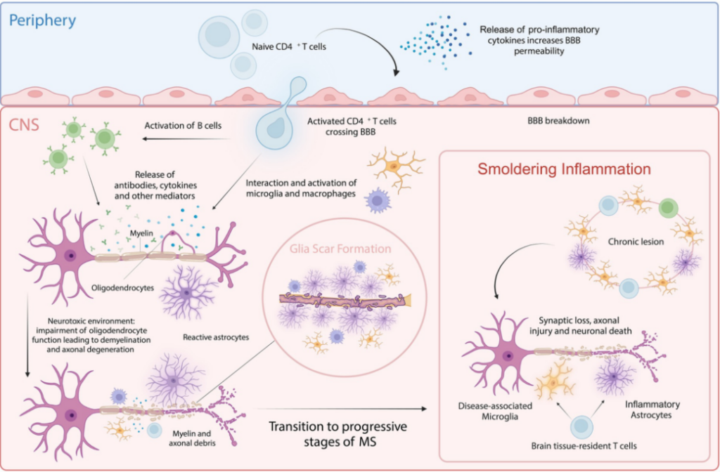
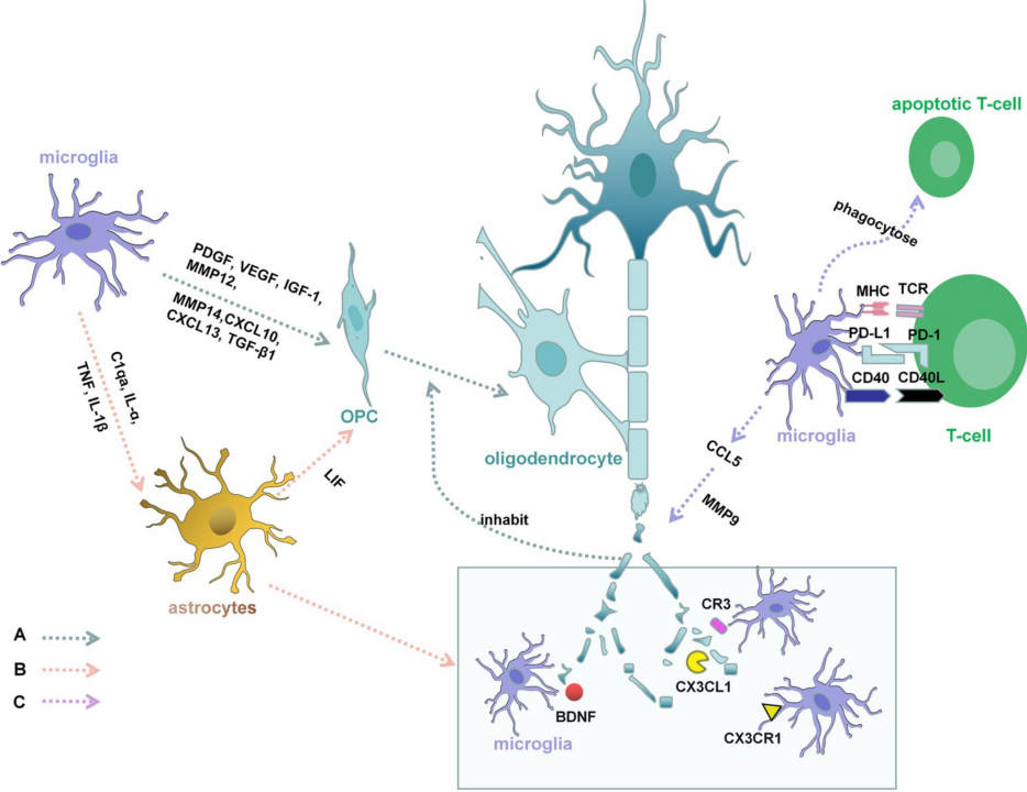
Within the chronic inflammatory microenvironment, microglia undergo sustained aberrant activation via TREM2-DAP12 and CX3CR1 signaling, polarizing into pro-inflammatory M1 or disease-associated microglia (DAM) phenotypes accompanied by dysregulated PI3K-Akt/mTOR metabolic pathways. These activated microglia continuously release neurotoxic mediators (TNF-α, IL-1β, reactive oxygen species/ROS) and GM-CSF to recruit additional peripheral immune cells, establishing a persistent inhibitory inflammatory niche that blocks remyelination mediated by oligodendrocyte precursor cell (OPC) differentiation. This also drives mitochondrial dysfunction, chronic axonal injury and irreversible neuronal loss, which underlies permanent neurological deficits and disease progression from RRMS toward SPMS or PPMS.
Preclinical Mouse Models for MS Research
- Prf1-/- Knockout Mice Global deletion of the perforin gene significantly alleviates clinical disease severity, with markedly reduced CNS inflammatory infiltration, demyelination and axonal damage.
- Tnf-/- Knockout Mice Loss of TNF-α diminishes central inflammatory responses, BBB breakdown, oligodendrocyte loss and demyelinating lesions.
- Il23p19-/- Knockout Mice Deletion of the IL-23 specific p19 subunit blunts pathogenic Th17 responses, lowering EAE incidence, delaying disease onset, reducing clinical severity and limiting inflammatory cell recruitment into the CNS.
- Cx3cr1-GFP Reporter Mice Green fluorescent protein (GFP) knocked into the microglia-specific Cx3cr1 locus enables real-time in vivo visualization of microglial morphology, distribution, migration and physical interactions with neurons and synapses under physiological, inflammatory (EAE) and neurodegenerative conditions.
VeloGene Biotechnology Accelerates Rare Disease Gene Therapy R&D
Gene therapy delivers transformative therapeutic prospects for rare immune and neurological disorders, yet preclinical mechanistic research and therapeutic efficacy validation are entirely dependent on standardized, well-characterized animal models.
Powered by our proprietary TurboMice™ technology, VeloGene Biotechnology has generated a comprehensive portfolio of rare disease gene-edited mouse models. TurboMice resolves two longstanding technical bottlenecks of traditional transgenic strain construction: lengthy breeding cycles and low success rates for complex multi-locus genetic modifications. The platform supports precise editing of nearly any target genomic locus and generates fully homozygous gene-edited mouse lines directly from embryonic stem cells in as little as 2 months.
VeloGene Biotechnology provides custom multiple sclerosis mouse models tailored to individual experimental requirements, including Prf1-/- knockout mice, Tnf-/- knockout mice, Il23p19-/- knockout mice and Cx3cr1-GFP reporter mice. Academic and industrial investigators are welcome to reach out for technical consultations!
References
[1] Wang Q, Lu ZQ, Li R. Advances in multiple sclerosis treatment. J Chongqing Med Univ. 2024, 49(5): 597-600. DOI:10.13406/j.cnki.cyxb.003500
[2] Patil MS, Lin LY, Marsh-Wakefield F, et al. Multiple Sclerosis: Immune Cells, Histopathology, and Therapeutics. Sclerosis. 2024, 2: 117-139. https://doi.org/10.3390/sclerosis2030009
[3] Zhang X, Chen F, Sun M, et al. Microglia in the context of multiple sclerosis. Front Neurol. 2023;14:1157287. doi:10.3389/fneur.2023.1157287
[4] Simões-Abade MBC, Patterer M, Nicaise AM, et al. Brain organoid methodologies to explore mechanisms of disease in progressive multiple sclerosis. Front Neurol. 2023;14:1157287. doi:10.3389/fneur.2023.1157287
Previous Article: Rare Diseases Focus | Issue 24: Hypophosphatemic Rickets
Next Article: Rare Diseases Focus | Issue 26: Tuberous Sclerosis Complex