




IgG4-related disease (IgG4-RD) is a chronic progressive fibrosing inflammatory disorder that received a unified international nomenclature in 2011. Capable of mimicking malignant tumors and involving nearly all visceral organs, it has become a core research focus in rheumatology and pathology.
It was officially included in China’s First National Rare Disease List in 2018. The condition predominantly affects males aged 50–70. Global epidemiological surveys report a prevalence of 0.5–1 case per 100,000 individuals. Due to highly variable clinical manifestations, misdiagnosis and missed diagnosis remain extremely common in clinical practice.
Pathogenesis
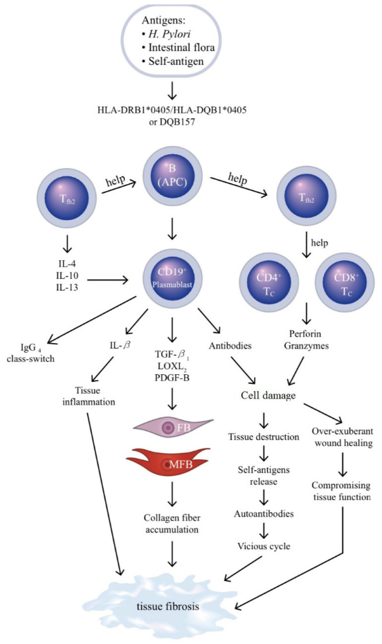
IgG4-RD arises from a dysregulated immune cascade driven by aberrantly activated T and B lymphocytes, ultimately leading to irreversible organ fibrosis.
Pathogenic cascade overview:
- Follicular helper T cells (Tfh) and cytotoxic CD4⁺ T cells (CD4-CTL) become hyperactivated and secrete large quantities of IL-4, IL-10, TGF-β and other cytokines.
- These cytokines drive two key pathological processes:
- Induce B cell differentiation into IgG4-producing plasma cells;
- Stimulate fibroblast-to-myofibroblast transition.
- Sustained TGF-β secretion acts as the primary driver of progressive tissue fibrosis, causing stiffening and loss of function in affected organs.
- Oligoclonal expansion of circulating CD19⁺ plasmablasts closely correlates with disease activity. This mechanism provides solid theoretical support for B cell-targeted biologics, fully delineating the pathological chain from immune dysregulation to permanent tissue damage.
Preclinical Mouse Models for IgG4-RD Research
IgG4-RD research models fall into two core categories: non-humanized strains (utilizing murine IgG1 as the functional ortholog of human IgG4) and humanized strains engineered to express human IgG4/human Fc fragments.
1. Non-Humanized Mouse Models (Murine IgG1 as IgG4 Ortholog)
Murine IgG1 shares highly homologous biological functions with human IgG4: both exhibit low Fcγ receptor affinity, weak complement activation capacity and intrinsic immunomodulatory/anti-inflammatory properties.
Researchers generate disease phenotypes via gene editing (LatY136F knock-in, CD28 knockout, Aire knockout, etc.) or active immunization to induce systemic Th2 skewing, polyclonal B cell activation and elevated serum IgG1.
Model Advantages
- Faithfully recapitulate hallmark IgG4-RD pathology: multi-organ lymphoplasmacytic infiltration, storiform fibrosis and obliterative phlebitis;
- Intact endogenous murine immune microenvironment without exogenous human protein introduction;
- Ideal for studying disease natural history, host genetic background and broad-spectrum anti-fibrotic drug screening.
2. Humanized IgG4-RD Mouse Models
These strains eliminate interspecies differences by reconstructing a humanized immune system expressing human IgG4. Construction approaches include transplanting patient-derived peripheral blood mononuclear cells (PBMC), CD34⁺ hematopoietic stem cells or genetically modified cell lines (K562, EBV-transformed B cells) into immunodeficient NSG mice.
Model Applications
- Visualize human IgG4⁺ plasma cell homing, tissue infiltration and germinal center formation in target organs (pancreas, salivary glands);
- Dynamically monitor circulating human IgG4 concentrations in vivo;
- Dissect the exact immunoregulatory mechanisms of human IgG4 Fc domains;
- Validate therapeutic antibodies targeting human IgG4 or human Fcγ receptors;
- Study synergistic pathogenic interactions between human CTLs and IgG4 under human physiological immune conditions.
VeloGene Biotechnology Accelerates IgG4-RD Research & Therapeutic Development
Novel biologic and gene therapies for rare fibro-inflammatory diseases rely entirely on well-characterized, precise animal models for preclinical validation.
Powered by proprietary TurboMice™ technology, VeloGene Biotechnology overcomes two historic limitations of traditional transgenic production: lengthy breeding cycles and low success rates for complex multi-locus gene edits. Our platform enables precise targeted editing of nearly any genomic locus. Fully homozygous gene-edited mouse lines can be generated directly from embryonic stem cells in as little as 2 months, eliminating time-consuming crossbreeding and screening workflows.
VeloGene Biotechnology provides custom IgG4-RD mouse strains including LatY136F knock-in mice and Aire knockout mice. Academic and industrial investigators are welcome to submit project consultations!
References
- Hsieh SC, Shen CY, Liao HT, et al. The Cellular and Molecular Bases of Allergy, Inflammation and Tissue Fibrosis in Patients with IgG4-related Disease. Int J Mol Sci. 2020;21(14):5082. DOI:10.3390/ijms21145082. PMID:32708432; PMCID:PMC7404109
- Yamada K, Zuka M, Ito K, et al. LatY136F knock-in mouse model for human IgG4-related disease. PLoS One. 2018;13(6):e0198417. DOI:10.1371/journal.pone.0198417. PMID:29902238; PMCID:PMC6002065
- Umehara H, Okazaki K, Kawa S, et al. The 2020 revised comprehensive diagnostic (RCD) criteria for IgG4-RD. Mod Rheumatol. 2021;31(3):529-533. DOI:10.1080/14397595. PMID:33274670; PMCID omitted