




What Is Hypophosphatemic Rickets?
Hypophosphatemic Rickets (HR) is a rare skeletal disorder with an incidence of approximately 1 in 25,000. It arises from genetic or acquired triggers that induce excessive renal phosphate excretion, leading to severe hypophosphatemia and impaired bone mineralization.
Clinical manifestations differ by age group:
- Children: Cranial bossing, pectus carinatum, rachitic rosary, bowleg/knock-knee limb deformities, growth retardation, short stature, waddling gait, and dental anomalies including enamel erosion and recurrent dental abscesses.
- Adults: Persistent bone pain, muscle weakness, multiple fractures, loss of height, and limited mobility.
Pathogenesis
Hypophosphatemic rickets is categorized into hereditary and acquired subtypes.
1. Hereditary Forms
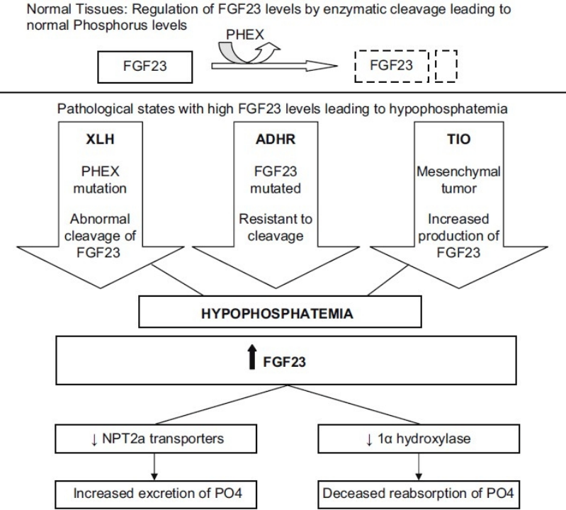
- X-linked Hypophosphatemia (XLH) The most prevalent hereditary subtype, accounting for 80% of cases, caused by loss-of-function variants in the PHEX gene.
- Autosomal Dominant Hypophosphatemic Rickets (ADHR) Driven by gain-of-function mutations in FGF23, with c.527G>A (p.Arg176Gln) as the most common pathogenic variant.
- Autosomal Recessive Hypophosphatemic Rickets (ARHR) Includes subtypes caused by mutations in DMP1, ENPP1, and FAM20C.
- Hereditary Hypophosphatemic Rickets with Hypercalciuria (HHRH) Resulting from pathogenic variants in the SLC34A3 gene.
2. Acquired Form
Tumor-Induced Osteomalacia (TIO)
Benign mesenchymal tumors overproduce intact bioactive FGF23, triggering systemic phosphate wasting.
Core Molecular Mechanism: FGF23-PHEX Axis Dysregulation
Fibroblast growth factor 23 (FGF23), secreted by osteocytes and osteoblasts, acts as the master endocrine regulator of phosphate homeostasis. The PHEX gene maps to Xp22.1 and is predominantly expressed in bone-forming cells.
Under physiological conditions, PHEX interacts with SIBLING family glycoproteins such as DMP1 to stabilize transcriptional repressor complexes that suppress FGF23 transcription, while preventing release of mineral-inhibiting ASARM peptides.
When PHEX loses function due to mutation:
- Uninhibited ASARM peptides accumulate to directly suppress bone mineralization and upregulate FGF23 expression;
- Osteocyte FGF23 transcription and secretion surge drastically.
Elevated circulating FGF23 downregulates serum 1,25-dihydroxyvitamin D, inhibits phosphate reabsorption in renal proximal tubules and reduces intestinal phosphate uptake, resulting in persistent hypophosphatemia and the full spectrum of rachitic/osteomalacic bone lesions.
Gene Therapy Strategies
1. Minicircle DNA (MC-DNA) Therapy
In October 2025, a research team led by Xu Chao and Zhao Jiajun at Shandong Provincial Hospital published work in Advanced Science. They delivered minicircle DNA encoding the FGF23 inhibitory fragment (amino acids 180–251) to XLH model mice. Treatment significantly restored serum phosphate levels, reduced circulating alkaline phosphatase, and rescued defective bone mineralization, with no obvious adverse effects observed over at least six weeks post-administration.
2. Liver-Targeted AAV Gene Therapy
Hepatic AAV vectors encoding the C-terminal inhibitory domain of FGF23 effectively ameliorate skeletal lesions and osteomalacia in XLH preclinical mouse models.
Preclinical Mouse Models for HR Research
- Dmp1<sup>-/-</sup> Knockout Mice Recapitulate ARHR1 pathology driven by DMP1 ablation, featuring markedly elevated FGF23, severe hypophosphatemia and global mineralization defects.
- Slc34a3<sup>-/-</sup> Knockout Mice Model HHRH, presenting disrupted phosphate homeostasis and characteristic skeletal lesions.
- Hyp Mutant Mice Classic well-established XLH research strain carrying endogenous Phex mutation, faithfully recapitulating human FGF23 overexpression and all core hypophosphatemic skeletal phenotypes.
- Phex T1349C Knock-In Mice Engineered to carry the human pathogenic T1349C variant in the murine Phex locus for genotype-specific XLH mechanistic studies.
- Fgf23 R176Q Knock-In Mice Harbor the ADHR hotspot p.Arg176Gln mutation, leading to constitutive FGF23 gain-of-function and disrupted phosphate metabolism with severe bone pathology.
VeloGene Biotechnology Accelerates Rare Disease Gene Therapy R&D
Gene therapy delivers transformative therapeutic prospects for rare inherited skeletal disorders, yet preclinical mechanism research and therapeutic efficacy validation are fully dependent on standardized, well-characterized animal models.
Powered by our proprietary TurboMice™ technology, VeloGene Biotechnology has developed an extensive portfolio of rare disease gene-edited mouse models. TurboMice™ resolves two longstanding technical bottlenecks of traditional transgenic strain construction: prolonged breeding cycles and low success rates for complex multi-locus genetic modifications. The platform supports precise editing of nearly any target genomic locus and generates fully homozygous gene-edited mouse lines directly from embryonic stem cells in as little as 2 months.
VeloGene Biotechnology provides custom hypophosphatemic rickets mouse models tailored to experimental requirements, including Dmp1<sup>-/-</sup> knockout mice, Slc34a3<sup>-/-</sup> knockout mice, Hyp mutant mice, Phex T1349C knock-in mice, and Fgf23 R176Q knock-in mice. Academic and industrial investigators are welcome to contact our technical team for project consultations!
References
[1] Ding GX. New advances in hypophosphatemic rickets research. Chin J Pract Clin Pediatr. 2019, 34(17): 1304-1308. DOI:10.3760/cma.j.issn.2095-428X.2019.17.006
[2] Jagtap VS, Sarathi V, Lila AR, et al. Hypophosphatemic rickets. Indian J Endocrinol Metab. 2012;16(2):177-182. DOI:10.4103/2230-8210.93733. PMID:22470852; PMCID:PMC3313733
[3] Lorenz-Depiereux B, Benet-Pages A, Eckstein G, et al. Hereditary hypophosphatemic rickets with hypercalciuria is caused by mutations in the sodium-phosphate cotransporter gene SLC34A3. Am J Hum Genet. 2006;78(2):193-201. DOI:10.1086/499410. PMID:16358215; PMCID:PMC1380229
[4] Jagtap VS, Sarathi V, Lila AR, et al. Hypophosphatemic rickets. Indian J Endocrinol Metab. 2012;16(2):177-182. DOI:10.4103/2230-8210.93733. PMID:22470852; PMCID:PMC3313733
Previous Article: Rare Diseases Focus | Issue 23: Pulmonary Alveolar Proteinosis
Next Article: Rare Diseases Focus | Issue 25: Multiple Sclerosis