



What Is Glycogen Storage Disease (Type I & Type II)?
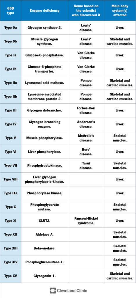
Glycogen Storage Disease (GSD) is a rare autosomal recessive metabolic disorder triggered by deficiencies in enzymes involved in glycogen synthesis or breakdown, resulting in abnormal glycogen accumulation in the liver, skeletal muscle and other visceral organs. At least 19 subtypes have been identified, among which Type I (hepatic form, von Gierke disease) and Type II (Pompe disease) are the most prevalent, jointly accounting for approximately 40% of all GSD cases.
- The incidence of GSD Type I ranges from 1 in 100,000 to 1 in 20,000 individuals;
- The incidence of GSD Type II ranges from 1 in 100,000 to 1 in 14,000 individuals, with slight variations across ethnic populations.
Subtypes of GSD Type I
- Type Ia (80% of all Type I cases) Caused by deficiency of glucose-6-phosphatase (G6PC), presenting the classic pentad: hypoglycemia, hepatomegaly, hyperlactatemia, hyperuricemia and hypertriglyceridemia.
- Type Ib (20% of all Type I cases) Caused by defects in the glucose-6-phosphate transporter (SLC37A4). Patients exhibit identical metabolic abnormalities to Type Ia, plus neutropenia and recurrent bacterial infections.
GSD Type II (Pompe Disease)
Accounting for 15% of total GSD cases, Pompe disease arises from deficiency of lysosomal acid α-glucosidase (GAA), leading to systemic glycogen deposition in muscle tissues. The infantile subtype manifests with hypertrophic cardiomyopathy and respiratory failure, while the late-onset subtype features progressive skeletal muscle weakness. It is clinically categorized into Infantile-Onset Pompe Disease (IOPD) and Late-Onset Pompe Disease (LOPD) based on age of onset and primary affected organs.
Pathogenesis
GSD Type Ia
Pathogenic variants in the G6PC gene induce loss of functional glucose-6-phosphatase-α. Glucose-6-phosphate (G6P) cannot be hydrolyzed into free glucose within the endoplasmic reticulum lumen, disrupting glycogenolysis and gluconeogenesis pathways. Intracellular G6P accumulates in hepatocytes, boosting glycolysis and triggering lactic acidosis; overactivation of the pentose phosphate pathway elevates uric acid synthesis; excess acetyl-CoA drives hyperlipidemia, collectively generating the characteristic pentad clinical profile of GSD Ia.
The two most prevalent pathogenic variants in Chinese patients are c.648G>T (57% allele frequency) and c.248G>A (14% allele frequency).
Figure source: PubMed
GSD Type Ib
Mutations in the SLC37A4 gene impair glucose-6-phosphate transporter (G6PT) function, blocking G6P translocation from the cytoplasm into the ER lumen for hydrolysis by G6Pase, thereby reproducing identical metabolic dysregulations as Type Ia. Meanwhile, loss of ER antioxidant protection in granulocytes accelerates neutrophil apoptosis and immune dysfunction.
The most common variants among Chinese patients are c.572C>T and c.446G>A.

GSD Type II (Pompe Disease)
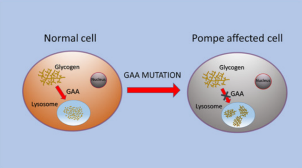
The causal GAA gene is located at chromosomal locus 17q25.3, containing 20 exons. Pathogenic mutations reduce lysosomal acid α-glucosidase activity, preventing glycogen degradation within lysosomes. Glycogen massively accumulates in lysosomes of skeletal muscle, cardiac muscle and smooth muscle cells, forming characteristic “glycogen vacuoles”. This triggers vacuolar degeneration of myocytes, disrupted autophagy-mitochondrial crosstalk and ultimately irreversible cell death.
Gene Therapy Strategies
1. Recombinant AAV Vector Therapy for GSD Type Ia
DTX401, an investigational therapy developed by Ultragenyx Pharmaceuticals, delivers AAV8-hG6PC via a single intravenous infusion. This vector efficiently delivers human G6PC cDNA to hepatocytes and drives sustained transgene expression under the native promoter, restoring G6Pase-α activity and reconstituting hepatic glucose output. Phase 3 clinical data confirmed that DTX401 reduces daily cornstarch supplementation and improves systemic metabolic biomarkers.
2. Somatic Gene Therapy for GSD Type Ib
Recombinant adeno-associated virus (rAAV) vectors are used to deliver human G6PT transgenes specifically to hepatocyte nuclei. The exogenous transgenes exist as episomal DNA without genomic integration, preserving the native genetic background of hepatocytes and conferring an excellent safety profile.
3. AAV-Mediated Gene Delivery for GSD Type II
Systemic administration of AAV9 or AAV8 vectors carrying the GAA transgene (controlled by dual muscle/liver-specific promoters) enables persistent secretion of functional acid α-glucosidase in the heart, skeletal muscle and central nervous system, clearing accumulated lysosomal glycogen. Both infantile and late-onset Pompe disease programs have entered Phase I/II clinical trials.
Preclinical Mouse Models for GSD Research
I. Models for GSD Type Ia
- G6pc-/- Knockout Mice High pre-weaning mortality; massive glycogen and lipid deposition in liver and kidneys, accompanied by severe hyperlactatemia.
- Alb-Cre G6pcflox/flox Conditional Knockout Mice Spontaneously develop hepatic adenomas and hepatocellular carcinoma, ideal for long-term toxicity and liver tumor mechanism research. G6Pase-α is selectively ablated only in hepatocytes, resulting in mild fasting hypoglycemia without continuous glucose supplementation requirements and largely preserved renal metabolic parameters.
- G6pc-R83C Point Knock-In Mice Recapitulates the prevalent human pathogenic variant, presenting disrupted glucose homeostasis and abolished hepatic G6Pase-α enzymatic activity.
II. Models for GSD Type Ib
- G6pt-/- Knockout Mice Reproduce core metabolic phenotypes of Type Ia including hypoglycemia, hyperlactatemia and hyperlipidemia, with additional neutropenia, intestinal inflammation and accelerated severe renal fibrosis.
- SLC37A4 Mutant Knock-In Mice Exhibit impaired glycogen metabolic homeostasis consistent with human GSD Ib pathology.
III. Models for GSD Type II (Pompe Disease)
- GAA-/- Global Knockout Mice Complete loss of systemic GAA enzymatic activity, massive lysosomal glycogen deposition in cardiac and skeletal muscle, left ventricular hypertrophy and progressive diaphragmatic weakness.
- GAA/GYS1 Double Knockout Mice Genetic ablation of glycogen synthase 1 in muscle eliminates muscular glycogen synthesis, drastically alleviating glycogen storage burden and cardiac hypertrophy.
- GAA p.R484Q Knock-In Mice Progressive skeletal muscle weakness and declining respiratory function onset at 6 months of age, faithfully recapitulating human late-onset Pompe disease and suitable for long-term therapeutic efficacy evaluation.
VeloGene Biotechnology Empowers Rare Disease Gene Therapy R&D
Gene therapy brings transformative therapeutic prospects for rare inherited disorders, yet preclinical development and therapeutic validation are fully dependent on standardized, well-characterized animal models.
Powered by our proprietary TurboMice™ platform, VeloGene Biotechnology has generated a comprehensive portfolio of rare disease gene-edited mouse models. TurboMice™ resolves two long-standing technical limitations of traditional transgenic model construction: prolonged breeding cycles and extremely low success rates for complex multi-locus gene modifications. This platform supports precise editing of nearly any target genomic locus and generates fully homozygous gene-edited mouse lines directly from embryonic stem cells in as little as 2 months.
VeloGene Biotechnology provides custom GSD Type I & Type II mouse models tailored to client research specifications, including G6pc<-/- mice, Alb-Cre G6pcflox/flox conditional knockout mice, G6pc-R83C point knock-in mice, G6pt-/- mice, SLC37A4 mutant knock-in mice, GAA-/- global knockout mice, GAA/GYS1 double knockout mice, and GAA p.R484Q knock-in mice. Academic and industrial researchers are welcome to contact our technical team for project consultations!
References
[1] Jiang JJ, Ma MS. Research progress and therapeutic advances in glycogen storage disease type Ib. J Rare Dis Res. 2024, 3(4): 522-526. DOI: 10.12376/i.issn.2097-0501.2024.04.016.
[2] Chou JY, Jun HS, Mansfield BC. Glycogen storage disease type I and G6Pase-β deficiency: etiology and therapy. Nat Rev Endocrinol. 2010;6(12):676-688. DOI: 10.1038/nrendo.2010.189. PMID: 20975743; PMCID: PMC4178929.
[3] Dan L, Song X, Yu H. A case of glycogen storage disease type Ia with gout as the initial manifestation. J Zhejiang Univ (Med Sci). 2023;52(2):230-236. DOI: 10.3724/zdxbyxb-2022-0530. PMID: 37283108; PMCID: PMC10409914.
[4] Raben N, Nagaraju K, Lee E, et al. Targeted disruption of the acid alpha-glucosidase gene in mice causes an illness with critical features of both infantile and adult human glycogen storage disease type II. J Biol Chem. 1998;273(30):19086-19092. DOI: 10.1074/jbc.273.30.19086. PMID: 9668092.
[5] Weinstein DA, Derks TG, Rodriguez-Buritica DF, et al. Safety and efficacy of DTX401, an AAV8-mediated liver-directed gene therapy, in adults with glycogen storage disease type Ia (GSDIa). J Inherit Metab Dis. 2025;48(2):e70014. DOI: 10.1002/jimd.70014. PMID: 40064185; PMCID: PMC11893205.
[6] Xie Y, Hu B, Gao Y, et al. Yap signalling regulates ductular reactions in CRISPR/Cas9-induced glycogen storage disease type Ia mice. Anim Cells Syst. 2022;26(6):300-309. DOI: 10.1080/19768354.2022.2139755. PMID: 36605584; PMCID: PMC9809376.
[7] Arnaoutova I, Aratyn-Schaus Y, Zhang L, et al. Base-editing corrects metabolic abnormalities in a humanized mouse model for glycogen storage disease type-Ia. Nat Commun. 2024;15(1):9729. DOI: 10.1038/s41467-024-54108-1. PMID: 39523369; PMCID: PMC11551175.