




What Is Fanconi Anemia?
Fanconi Anemia (FA) is a rare inherited bone marrow failure syndrome. Its primary inheritance mode is autosomal recessive, while approximately 2% of cases show X-linked recessive transmission. The disease is characterized by congenital developmental abnormalities, progressive bone marrow failure, and drastically elevated cancer susceptibility. Common clinical manifestations include café-au-lait spots (55%), short stature (51%), limb malformations (43%), craniofacial anomalies (26%), ocular defects (23%) and renal abnormalities.
The global incidence stands at roughly 1 in 160,000, with a lower prevalence in Asian populations. The cumulative cancer risk across all FA patients reaches around 30%, encompassing hematological malignancies such as myelodysplastic syndrome (MDS) and acute myeloid leukemia, as well as solid tumors including head and neck squamous cell carcinoma and breast/ovarian cancer; solid tumors account for 30%–40% of all malignant cases.
Pathogenesis
The core pathogenic driver of FA is impaired function of the FA/BRCA DNA repair pathway, which abolishes cellular capacity to resolve DNA interstrand crosslinks (ICLs). ICLs represent severe covalent lesions bridging the two strands of DNA, physically obstructing DNA replication and transcription and inducing genomic instability.
From a molecular epidemiological perspective, FANCA mutations predominate among FA patients (60%–70%), followed by FANCC (7%–15%) and FANCG (~10%).
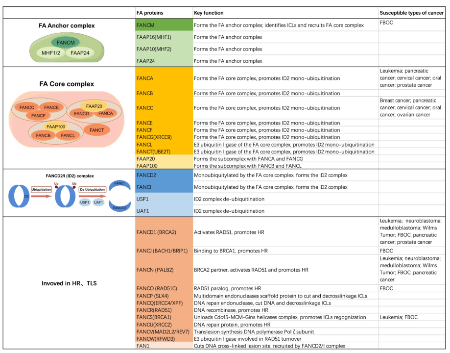
When replication forks stall upon encountering ICLs, the FANCM–FAAP24–MHF1/2 complex recognizes and binds the stalled fork, then recruits the FA core complex composed of at least eight proteins including FANCA, FANCB and FANCC. Within this core complex, the E3 ubiquitin ligase FANCL catalyzes monoubiquitination of the FANCD2–FANCI heterodimer (ID2 complex), serving as a molecular switch to activate the entire FA pathway. Monoubiquitinated ID2 dissociates from the core complex and tightly associates with chromatin adjacent to ICL lesions, acting as a scaffold to recruit downstream repair effectors such as FANCP/SLX4 and the XPF–ERCC1 (FANCQ) endonuclease for crosslink unhooking and cleavage.
After crosslink incision, DNA gap repair proceeds via two coordinated pathways:
- Translesion Synthesis (TLS): Error-prone polymerases including REV1 and Polζ insert nucleotides opposite the damaged template strand;
- Homologous Recombination (HR): High-fidelity repair utilizing the sister chromatid as a template, mediated by the BRCA2 (FANCD1)–PALB2 (FANCN)–RAD51 (FANCR) complex to execute strand invasion and Holliday junction resolution.
Once repair is complete, the USP1–UAF1 deubiquitinase complex removes ubiquitin moieties from FANCD2 to terminate pathway activation.
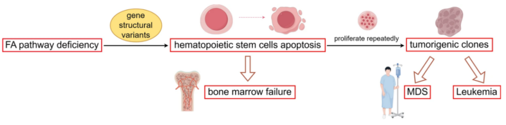
Biallelic loss-of-function mutations in any FA gene disrupt the entire repair cascade. Cells lose the ability to eliminate endogenous (e.g., aldehyde metabolites) or exogenous (chemotherapeutic agents) ICL damage, leading to accumulated DNA lesions, chromosomal breakage, and ultimately progressive bone marrow failure, congenital malformations and drastically elevated tumor predisposition such as acute myeloid leukemia and head-neck squamous carcinoma.
Gene Therapy
RP-L102 Lentiviral Gene Therapy
Self-inactivating lentiviral vectors deliver functional wild-type FANCA into patient-derived autologous CD34+ hematopoietic stem cells to restore physiological DNA crosslink repair capacity. After mobilization with G-CSF and plerixafor, harvested stem cells undergo ex vivo transduction under optimized low-oxygen short-term culture conditions. This therapeutic candidate has entered multi-center Phase II clinical trials.
Preclinical Mouse Models for FA Research
- Usp1-/- Knockout Mice Global ablation of the Usp1 gene results in persistent monoubiquitination of FANCD2, disrupting nuclear foci formation and ICL repair efficiency. Phenotypes include hematopoietic defects and germ cell depletion, with extreme hypersensitivity to crosslinking agents. This strain is an ideal model to investigate negative regulatory mechanisms and deubiquitination within the FA pathway.
- Fanca-/- Knockout Mice Targeted deletion of exons 4–7 in the murine Fanca gene. These animals display no obvious congenital or severe hematological defects yet present markedly reduced fertility, widely utilized to dissect core FA complex function.
- Fancc-/- Knockout Mice Exhibit profound hypersensitivity to DNA crosslinkers such as mitomycin C (MMC) accompanied by increased chromosomal fragmentation, a key tool for studying early FA pathway activation events.
- Fancd2-/- Knockout Mice Deletion of the central pathway effector FANCD2 abolishes all downstream DNA repair functions. The model presents severe phenotypes with high embryonic lethality; surviving animals display prominent developmental anomalies, bone marrow failure and heightened cancer susceptibility, serving as the classic preclinical strain to interrogate core FA pathway mechanisms and hematopoietic stem cell biology.
VeloGene Biotechnology Accelerates Rare Disease Gene Therapy R&D
Gene therapy delivers transformative therapeutic prospects for rare inherited hematological disorders, yet preclinical mechanistic research and therapeutic efficacy validation fully rely on standardized, well-characterized animal models.
Powered by our proprietary TurboMice™ technology, VeloGene Biotechnology resolves two longstanding technical bottlenecks of traditional transgenic strain construction: prolonged breeding cycles and low success rates for complex multi-locus genetic modifications. The platform supports precise editing of nearly any target genomic locus and generates fully homozygous gene-edited mouse lines directly from embryonic stem cells in as little as 2 months.
VeloGene Biotechnology provides custom Fanconi anemia mouse models including Usp1-/-, Fanca-/-, Fancc-/-, and Fancd2-/- knockout mice. Academic and industrial investigators are welcome to contact our technical team for project consultations!