




In 2024, major pharmaceutical corporations heavily invested in the TIGIT (T cell immunoreceptor with Ig and ITIM domains) target within the tumor immunotherapy space. Despite setbacks in several Phase III clinical trials, encouraging clinical data from the combination regimen of tiragolumab (anti-TIGIT monoclonal antibody) and atezolizumab (anti-PD-L1 antibody for PD-L1-high non-small cell lung cancer) has reignished global industry optimism for TIGIT as a next-generation immune checkpoint inhibitor.
With its unique dual immunomodulatory mechanisms, TIGIT has emerged as a star target in tumor immunotherapy. Let’s dive deep into its biological functions.
Introduction to the TIGIT Target
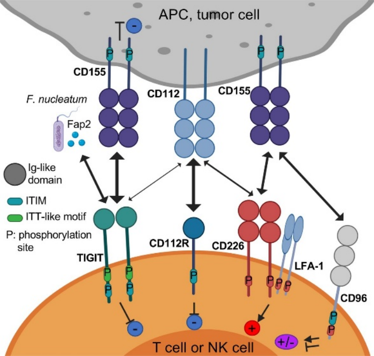
TIGIT is an immune checkpoint protein predominantly expressed on the surface of T cells and natural killer (NK) cells. Belonging to the immunoglobulin superfamily, its structure consists of an extracellular IgV (immunoglobulin variable) domain, a transmembrane segment, and an intracellular cytoplasmic tail carrying ITIM (immunoreceptor tyrosine-based inhibitory) and ITT (immunoglobulin tyrosine tail) motifs. This structural architecture enables TIGIT to transmit inhibitory immune signals. By binding with high affinity to ligands including CD155 (PVR), TIGIT acts as a key immune checkpoint within the tumor microenvironment.
I. Three Core Inhibitory Mechanisms of TIGIT Mediating Tumor Immune Evasion
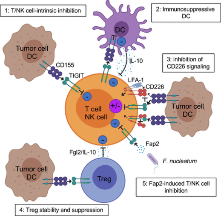
TIGIT suppresses immune cell function via multiple pathways to facilitate tumor immune escape, with three primary modes of action:
- Effects on dendritic cells (DCs) Upon TIGIT-CD155 ligation on DCs, inhibitory signaling is triggered within CD155 itself, hindering DC maturation and activation, blunting antigen presentation capacity, and ultimately blocking effective T cell priming.
- Direct suppression of T and NK cells When TIGIT engages CD155 on T/NK cells, intracellular ITIM and ITT motifs recruit and activate phosphatases such as SHP1/2. These enzymes dephosphorylate and suppress critical signaling molecules including CD3ζ and ZAP70, directly inhibiting T cell and NK cell activation and cytotoxicity.
- Competitive antagonism of co-stimulatory receptor CD226 TIGIT binds CD155 with higher affinity than the co-stimulatory receptor CD226, competitively occupying ligand binding sites and ablating CD226-mediated T/NK cell activating signals. Figure Source: TIGIT in cancer immunotherapy
II. Complex Regulatory Roles of TIGIT in Autoimmune Diseases
1. Rheumatoid Arthritis (RA)
Reduced TIGIT expression on CD4⁺ T cells correlates positively with RA disease activity. TIGIT overexpression suppresses secretion of pro-inflammatory cytokines IFN-γ and IL-10 while boosting anti-inflammatory IL-10, alleviating arthritis pathology in preclinical models. However, TIGIT exerts opposing effects on follicular helper T (Tfh) cells: it upregulates anti-apoptotic proteins such as Bcl-xL to prolong Tfh survival, amplifying B cell activation and autoantibody production. On NK cells, TIGIT functions as a negative regulator; its expression is markedly downregulated in RA patients, leading to excessive NK cell activation and aggravated inflammation.
2. Systemic Lupus Erythematosus (SLE)
Elevated TIGIT levels on CD4⁺ T cells correlate with SLE disease activity. TIGIT-CD15 ligation negatively constrains CD4⁺ T cell proliferation and IFN-γ release, conferring protective effects. Its activity varies across T cell subsets: TIGIT enhances the helper function of Tfh cells, whereas high TIGIT expression on follicular regulatory T (Tfr) cells restrains autoantibody generation. NK cells from SLE patients show significantly diminished TIGIT expression; blockade of the TIGIT axis restores NK activity, confirming persistent TIGIT-mediated NK suppression.
3. Inflammatory Bowel Disease (IBD)
Mucosal CD4⁺ and CD8⁺ T cells from active IBD patients exhibit drastically reduced TIGIT expression. TIGIT serves as an activation marker for regulatory T cells (Tregs) and strengthens their immunosuppressive capacity; enhanced TIGIT signaling alleviates inflammation in chronic colitis models. Mechanistically, CD155 on DCs binds TIGIT on Tregs to curtail pro-inflammatory IL-12 secretion. Conversely, NK cells from IBD patients display elevated TIGIT expression, and TIGIT inhibition restores NK cytotoxic function.
Preclinical Mouse Models for TIGIT Research
- **Tigit<sup>-/-</sup> Knockout Mice Used to investigate the fundamental impacts of global TIGIT deletion on systemic immunity and anti-tumor immune responses.
- Humanized TIGIT Mice Endogenous murine Tigit locus replaced with full-length human TIGIT gene. Ideal for in vivo pharmacodynamic and safety evaluation of human-specific anti-TIGIT monoclonal antibodies.
- Dual Humanized hTIGIT/hPD-L1 Mice Simultaneously express human TIGIT and human PD-L1; a dedicated model for precise efficacy assessment of combined TIGIT and PD-1/PD-L1 inhibitor regimens.
VeloGene Biotechnology Accelerates Immunology Mechanism & Drug R&D
Gene and immunotherapies bring promising therapeutic options for common and autoimmune disorders, yet preclinical research and efficacy validation are fully dependent on standardized, well-characterized animal models.
Powered by proprietary TurboMice™ technology, VeloGene Biotechnology resolves two longstanding technical pain points of traditional transgenic strain construction: lengthy breeding cycles and low success rates for complex multi-locus gene edits. Our platform enables precise targeting of nearly any genomic locus, generating fully homozygous gene-edited mouse lines directly from embryonic stem cells in as little as two months.
VeloGene Biotechnology provides customized TIGIT-related mouse strains including Tigit<sup>-/-</sup> knockout mice, humanized TIGIT mice, and dual hTIGIT/hPD-L1 humanized mice. Academic and industrial researchers are welcome to reach out for project consultations!
References
[1] Chauvin J, Zarour HM. TIGIT in cancer immunotherapy. JITC. 2020;8:e000957. https://doi.org/10.1136/jitc-2020-000957
[2] Yue C, Gao S, Li S, et al. TIGIT as a Promising Therapeutic Target in Autoimmune Diseases. Front Immunol. 2022;13:911919. DOI:10.3389/fimmu.2022.911919. PMID: 35720417; PMCID: PMC9203892