




What Is Spinocerebellar Ataxia?
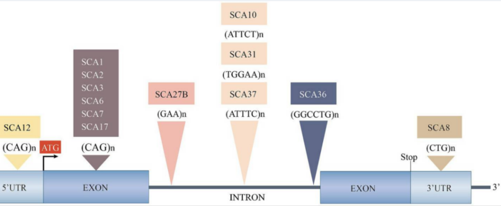
Spinocerebellar Ataxia (SCA) refers to a heterogeneous group of inherited neurodegenerative disorders triggered by distinct pathogenic gene mutations. Many SCA subtypes are classified as dynamic repeat expansion diseases, predominantly driven by expanded CAG trinucleotide repeats.
Core clinical manifestations include progressively unsteady gait, limb incoordination, dysarthria and abnormal ocular movements. Additional neurological complications observed in different subtypes may encompass peripheral neuropathy, cognitive decline and extrapyramidal signs. The global incidence ranges from 1 to 5 cases per 100,000 individuals. Among Chinese SCA patients, SCA3 represents the most prevalent subtype, accounting for 51.1% to 72.5% of all cases.
To date, more than 40 SCA subtypes (SCA1–SCA40) have been identified and categorized based on their causal genes. Their primary genetic mechanisms fall into two major groups:
- Polyglutamine (polyQ) disorders caused by expanded CAG repeats within coding regions (e.g., SCA1, SCA2, SCA3, SCA6, SCA7);
- Non-polyQ SCAs induced by non-coding repeat expansions or point mutations (e.g., SCA8, SCA10, SCA12, SCA31).
Pathogenesis
The pathogenic mechanisms of SCAs are grouped into three core molecular cascades based on distinct disease-driving genes:
1. PolyQ Protein Toxicity Pathway
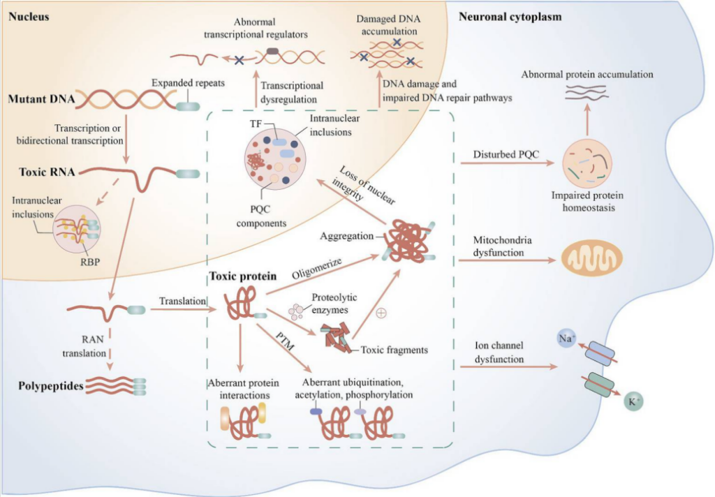
Driven by expanded CAG trinucleotide repeats in coding regions of genes including ATXN1, ATXN2, ATXN3, CACNA1A, and ATXN7. Mutant genes encode aberrantly elongated polyglutamine-containing proteins prone to misfolding and intranuclear aggregate formation. These toxic protein inclusions directly damage neurons and broadly disrupt cellular protein quality control (PQC) systems: they impair the ubiquitin-proteasome pathway, deplete molecular chaperones and inhibit autophagy-lysosomal clearance machinery. This triggers a cascade of pathological events including transcriptional dysregulation and mitochondrial dysfunction, ultimately leading to selective degeneration of cerebellar Purkinje cells and other neurons.
2. RNA Toxicity & RAN Translation Pathway
Triggered by expanded repeat sequences in non-coding loci such as ATXN8OS/ATXN8, ATXN10, PPP2R2B (SCA12), BEAN1/TK2 (SCA31), NOP56 (SCA36), and DAB1 (SCA37). Expanded repetitive DNA templates transcribe toxic RNA transcripts that accumulate as intranuclear RNA foci. These abnormal RNA clusters sequester essential RNA-binding proteins and disrupt physiological RNA homeostasis, including splicing, cytoplasmic transport and transcript stability. Furthermore, some expanded repeats undergo Repeat-Associated Non-AUG (RAN) translation to generate neurotoxic polypeptides (polyglutamine, polyalanine etc.), inflicting secondary proteotoxic injury. This pathway serves as the primary disease driver for SCA8, SCA10, SCA12, SCA31, SCA36 and SCA37.
3. Aberrant Native Protein Function Pathway
Caused by point mutations, small insertions or deletions in genes such as SPTBN2, TTBK2, PRKCG, TRPC3, KCND3, KCNC3, and FGF14, with missense variants predominating. Pathogenic alterations induce two main types of cellular dysfunction:
- Gain-of-function toxicity: mutant proteins misaggregate, exhibit hyperactive enzymatic activity or disrupt ion channel function (e.g., TRPC3, CACNA1A subunits);
- Loss-of-function/haploinsufficiency: impaired physiological protein activity disrupt signal transduction, kinase function and cytoskeletal stability.
All three pathological cascades converge to disrupt neuronal homeostasis and induce progressive loss of Purkinje cells and other vulnerable neurons, manifesting as worsening cerebellar ataxia and multi-system neurological impairment. Widespread breakdown of the cellular protein homeostasis network represents the shared terminal pathological hub across nearly all SCA subtypes.
Preclinical Mouse Models for SCA Research
- SCA1 Knock-In Mice Engineered via targeted insertion of expanded CAG repeats to faithfully recapitulate the natural disease progression of human SCA1. Widely utilized to investigate intranuclear inclusion formation and respiratory neural circuit dysfunction.
- SCA6 Knock-In Mice Generated by expanding endogenous Cacna1a CAG repeats in mice. These animals display prominent motor incoordination and serve as a vital tool for SCA6 pathogenic studies and long-term therapeutic efficacy assessment.
- SCA7 Knock-In Mice Created with expanded Atxn7 CAG repeats, recapitulating cerebellar degeneration accompanied by retinal atrophy; ideal for studying the multi-organ involvement unique to SCA7.
- Humanized SCA3 Mice Endogenous mouse Atxn3 is replaced with human ATXN3 carrying pathogenic CAG expansions, precisely modeling human SCA3 pathology and supporting screening of human-targeted nucleic acid therapeutics.
- SCA8 Mice Carry the human ATXN8OS/ATXN8 locus with expanded CTG•CAG repeats, recapitulating cerebellar dysfunction and polyglutamine/polyalanine aggregate formation; primarily used to dissect toxicity induced by non-coding repeat expansions.
- SCA41 Point Mutation Mice Engineered to harbor disease-causing missense variants in the Trpc3 gene, reproducing gait ataxia and Purkinje cell abnormalities for mechanistic research on ion channel-mediated spinocerebellar ataxia.
VeloGene Biotechnology Accelerates SCA Gene Therapy R&D
Gene therapy brings promising therapeutic prospects for rare hereditary neurodegenerative diseases, yet preclinical mechanistic research and therapeutic efficacy validation fully rely on standardized, well-characterized animal models.
Powered by our proprietary TurboMice™ technology, VeloGene Biotechnology resolves two longstanding technical bottlenecks of traditional transgenic strain construction: prolonged breeding cycles and low success rates for complex multi-locus genetic modifications. The platform supports precise editing of nearly any target genomic locus and generates fully homozygous gene-edited mouse lines in as little as 2 months directly from embryonic stem cells.
VeloGene Biotechnology provides custom spinocerebellar ataxia mouse models including SCA1 knock-in mice, SCA6 knock-in mice, SCA7 knock-in mice, humanized SCA3 mice, SCA8 mice and SCA41 point mutation mice. Academic and industrial investigators are welcome to contact our technical team for project consultations!
References
[1] https://www.ncbi.nlm.nih.gov/books/NBK557816/
[2] Cui ZT, et al. Spinocerebellar ataxias: from pathogenesis to recent therapeutic advances. Front Neurosci. 2024;18:1422442. DOI:10.3389/fnins.2024.1422442. PMID:38894941; PMCID:PMC11185097
[3] Garden GA, La Spada AR. Molecular pathogenesis and cellular pathology of spinocerebellar ataxia type 7 neurodegeneration. Cerebellum. 2008;7(2):138-149. DOI:10.1007/s12311-008-0027-y. PMID:18418675; PMCID:PMC4195584