




What Is Retinitis Pigmentosa?
Retinitis Pigmentosa (RP) is a highly genetically heterogeneous degenerative retinal disorder pathologically characterized progressive photoreceptor loss and retinal pigment epithelium (RPE) atrophy. Core clinical manifestations include nyctalopia, gradual peripheral visual field constriction, and progressive central vision loss. The classic fundus “triad” consists of bone-spicule pigment deposits, generalized retinal vascular attenuation, and waxy pallor of the optic disc. Multiple gene mutations and aberrant protein metabolism are confirmed as key drivers of RP pathogenesis.
RP is categorized into three major inheritance patterns: autosomal dominant RP (adRP, 15%–25%), autosomal recessive RP (arRP, 5%–20%), and X-linked RP (XLRP, 10%–15%). The global prevalence ranges from 1 in 3,000 to 1 in 7,000, while the domestic prevalence in China stands at approximately 1 in 4,000, affecting over 1.5 million individuals nationwide.
Pathogenesis
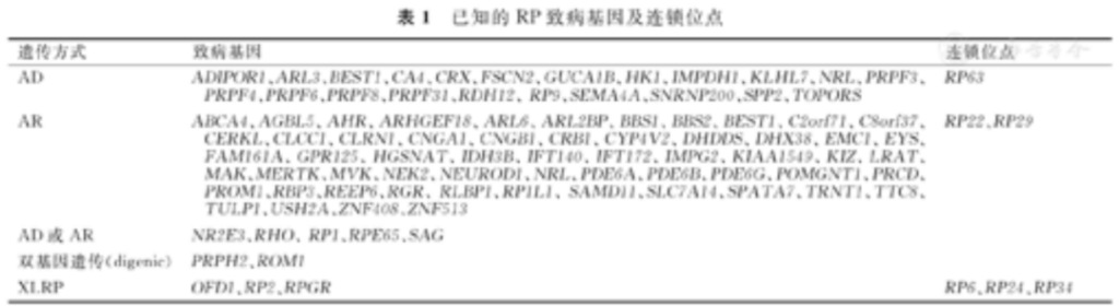
RP exhibits profound genetic heterogeneity; more than 90 causative genes have been identified to date, with point mutations as the predominant pathogenic variants. Its core pathological cascade is initiated by mutant photoreceptor-specific proteins (e.g., rhodopsin) with abnormal synthesis or impaired function, triggering sequential degenerative damage.
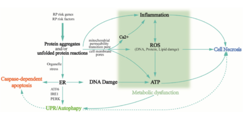
Pathogenic mutations trigger two primary initial injuries:
- Mutant misfolded proteins (e.g., mutated rhodopsin encoded by RHO) accumulate within the endoplasmic reticulum (ER), activating the unfolded protein response. Sustained unresolved ER stress induces calcium efflux and activates effector molecules such as caspase-12 to launch the intrinsic apoptotic pathway.
- Mutations in ciliary genes (e.g., RPGR) disrupt intraflagellar transport, impairing outer segment disc renewal and dysregulating cGMP and calcium homeostasis. Persistent high intracellular calcium activates calcium-dependent proteases and initiates apoptotic cascades.
Additionally, photoreceptors feature high oxygen consumption and outer segments rich in polyunsaturated fatty acids, rendering them highly susceptible to oxidative stress under genetic predisposition. Excessive reactive oxygen species (ROS) drive widespread lipid peroxidation, protein damage and DNA fragmentation, amplifying cellular injury and apoptotic signaling. Autophagy becomes dysregulated in this process: initially upregulated as an adaptive response, autophagic flux later stalls, leading to buildup of undegraded toxic substrates that further exacerbate cellular stress.
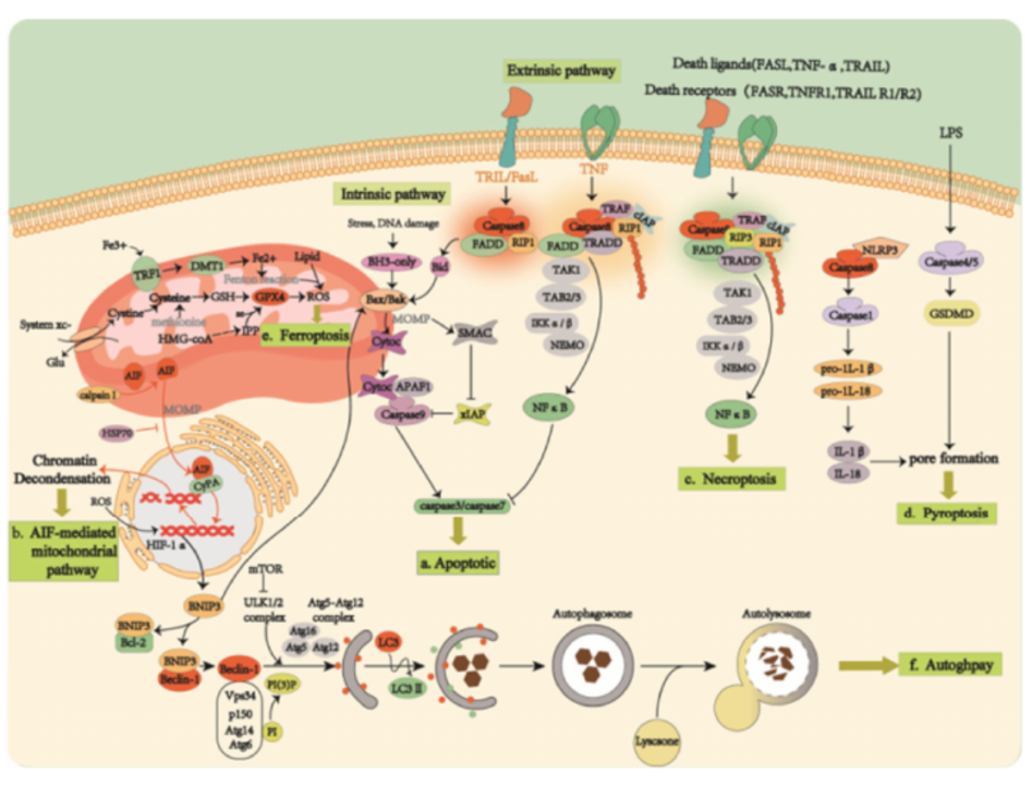
Damaged and dying photoreceptors release damage-associated molecular patterns (DAMPs) that activate pattern recognition receptors and propagate multiple cell death cascades via two major axes:
- DAMPs induce secretion of pro-inflammatory cytokines including TNF-α, which bind death receptors such as TNFR1 to trigger extrinsic apoptosis. If caspase-8 is suppressed, signaling shifts toward RIPK1/RIPK3/MLKL-mediated necroptosis.
- DAMPs such as ATP and crystalline substances directly activate NLRP3 inflammasomes, cleaving pro-GSDMD into active gasdermin D to induce pyroptosis with robust secretion of IL-1β and IL-18 that amplify inflammatory responses. Furthermore, impaired GPX4 function concurrent with oxidative stress drives lipid peroxidation accumulation and ferroptosis. Collectively, overlapping and synergistic signaling of apoptosis, necroptosis, pyroptosis and ferroptosis results in progressive loss of rod and cone photoreceptors.
Gene Therapy Strategies
1. Gene Replacement Therapy
Primarily applicable to autosomal recessive RP (arRP) and X-linked RP (XLRP). Recombinant adeno-associated viral (rAAV) vectors deliver intact wild-type genes to retinal cells to compensate for absent or dysfunctional proteins caused by pathogenic variants. Serotypes AAV2, AAV5, AAV8 and AAV9 are widely administered via intravitreal or subretinal injection. Luxturna® (voretigene neparvovec) has received marketing approval for RPE65-associated Leber Congenital Amaurosis. Multiple Phase 1 to Phase 3 clinical trials for XLRP (AAV5-RPGR) and PDE6B-deficient RP (AAV2/5-hPDE6B) are underway to slow photoreceptor degeneration.
2. Gene Silencing Technology
Designed for autosomal dominant RP (adRP) driven by dominant-negative mutations to suppress toxic mutant allele expression.
- RNA Interference (RNAi): Synthetic siRNA or miRNA targets mutant mRNA for degradation to reduce accumulation of toxic mutant proteins (e.g., certain RHO variants).
- Antisense Oligonucleotide (ASO): Modulates pre-mRNA splicing or accelerates mutant mRNA clearance to restore functional protein production. Approved ASO drugs such as Patisiran provide translational reference for RP therapeutics.
Preclinical Mouse Models for RP Research
- P23H-RHO Mice Recapitulates the most prevalent human rhodopsin missense variant Pro23His, characterized by progressive rod degeneration followed by secondary cone loss. Widely adopted for pathogenesis study, drug screening and gene therapy efficacy validation.
- S334ter-RHO Mice Carries a truncating RHO mutation deleting the C-terminal rhodopsin domain, presenting accelerated retinal degeneration: outer segment malformation appears at postnatal week 1, with nearly complete rod ablation by week 3.
- RHO-/- Knockout Mice Complete loss of rhodopsin expression, leading to defective rod development and absent outer segment formation with relatively slow apoptotic progression. Utilized to investigate rhodopsin’s physiological roles in photoreceptor maturation and function.
- RPGR-/- Knockout Mice Full depletion of RPGR protein; photoreceptors develop normally in early life but suffer disrupted outer segment turnover, with rod death initiating at 4–6 months followed by cone impairment. A gold-standard model for XLRP mechanistic research.
- RPGR<sup>rd9</sup> Mutant Mice Harbor exon 9 deletion in the murine Rpgr gene, displaying progressive rod dystrophy and reduced ERG amplitudes, ideal for defining therapeutic intervention windows.
VeloGene Biotechnology Accelerates Rare Disease Gene Therapy R&D
Gene therapy delivers transformative therapeutic prospects for rare inherited retinal degenerative disorders, yet preclinical mechanistic research and therapeutic efficacy validation fully rely on standardized, well-characterized animal models.
Powered by our proprietary TurboMice™ technology, VeloGene Biotechnology resolves two longstanding technical bottlenecks of traditional transgenic strain construction: prolonged breeding cycles and low success rates for complex multi-locus genetic modifications. The platform supports precise editing of nearly any target genomic locus and generates fully homozygous gene-edited mouse lines directly from embryonic stem cells in as little as 2 months.
VeloGene Biotechnology provides custom retinitis pigmentosa mouse models including P23H-RHO mice, S334ter-RHO mice, RHO-/- knockout mice, RPGR-/- knockout mice and RPGRrd9 mutant mice. Academic and industrial investigators are welcome to contact our technical team for project consultations!