




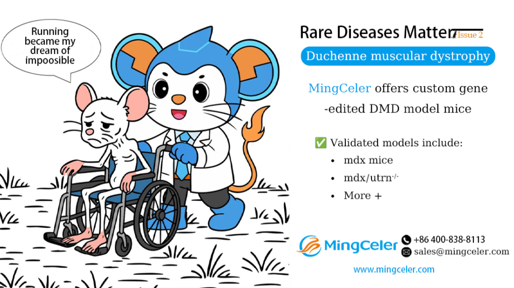
Duchenne muscular dystrophy (DMD) is a rare X-linked recessive genetic disorder caused by mutations in the DMD gene, leading to the loss of dystrophin protein. Dystrophin is critical for maintaining the stability of muscle cell membranes.
The incidence of DMD is approximately 1 in 3,500 to 5,000 live male births worldwide, with an estimated 450,000 to 600,000 patients globally. In China, the prevalence is about 8.5 per 100,000 males, with a total patient population of around 60,000.
DMD primarily affects skeletal and cardiac muscle. Patients typically present with a waddling gait, toe-walking, and lumbar lordosis. As the disease progresses, most patients lose the ability to walk by around age 12. Cardiac involvement can lead to dilated cardiomyopathy, conduction abnormalities, and arrhythmias. The average life expectancy is reduced to approximately 26 years.

Pathogenesis
The DMD gene is the largest known human gene, spanning 2.2 million base pairs with 79 exons. Mutations in this gene lead to the absence or dysfunction of dystrophin, destabilizing muscle cell membranes and causing progressive muscle fiber damage and degeneration.
Dystrophin is mainly expressed in skeletal muscle, cardiac muscle, and the brain, where it stabilizes the cytoskeleton and protects muscle cells.
Due to its large size, the DMD gene has a high mutation rate (approximately 1/10,000). Mutation types include:
- Deletion mutations: ~65%
- Duplication mutations: 6%–10%
- Point mutations, small insertions/deletions: 25%–30%
Deletion and duplication hotspots are located at the 5′ region (~20%) and central region (~80%) of the gene. Point mutations and small indels are scattered randomly.


Treatment Approaches
Currently, DMD is incurable, but advances in pathogenesis research and emerging technologies such as gene therapy and stem cell therapy are expanding treatment options.
1. Glucocorticoid Therapy
Glucocorticoids (e.g., prednisone) are the only standard treatment shown to improve muscle strength and slow disease progression by reducing inflammatory damage to muscle. However, they do not address the root genetic cause.
2. Stem Cell Therapy
Stem cell transplantation aims to regenerate muscle cells and restore dystrophin expression. Current research shows limited myogenic differentiation efficiency, and only a small number of bone marrow mesenchymal stem cells (BM-MSCs) contribute to new muscle fiber formation.
3. Gene Therapy
Five major gene therapy strategies are under development:
(1) Stop Codon Read-Through
Approximately 15% of DMD cases are caused by nonsense mutations (premature termination codons, PTCs). Aminoglycosides and other small molecules can induce ribosomal read-through of PTCs, allowing production of full-length dystrophin.
(2) Exon Skipping
Exon skipping uses antisense oligonucleotides (ASOs) to skip specific exons during pre-mRNA splicing, restoring the reading frame and producing a truncated but partially functional dystrophin.
(3) Exon Excision
Gene-editing nucleases (e.g., ZFNs) permanently remove critical splicing sequences in exon 51, preventing its transcription. This strategy can restore the open reading frame in about 13% of DMD patients.
(4) Gene Editing
Gene editing directly corrects the DMD mutation at the genomic DNA level, enabling permanent restoration of functional dystrophin under endogenous regulation. This approach offers the potential for one-time curative therapy.
(5) Micro-dystrophin Gene Therapy
Due to the huge size of the full DMD gene, viral delivery is challenging. Micro-dystrophin (a shortened, functional construct) is delivered via viral vectors to alleviate disease phenotypes. Multiple clinical trials are ongoing.
DMD Mouse Models
1. mdx Mouse
The mdx mouse carries a nonsense point mutation in exon 23 of the Dmd gene, resulting in the loss of full-length dystrophin. It is the most widely used DMD model.
However, its phenotype is milder than human DMD, with only a 20% reduction in lifespan and slow, fluctuating skeletal muscle lesions.
2. Double Knockout Mouse: mdx/utrn⁻/⁻
The mdx/utrn⁻/⁻ model lacks both dystrophin and utrophin, leading to severe, progressive muscular dystrophy with profound weakness, joint contractures, kyphosis, and a median survival of only ~3 months. This model closely recapitulates severe human DMD.
VeloGene Biotechnology Empowers DMD Gene Therapy R&D
Gene therapy for rare diseases relies on validated animal models.
Using our proprietary TurboMice™ technology, MingCeler Bio has developed a series of rare disease mouse models.
TurboMice™ overcomes the long timelines and low success rates of conventional methods, enabling in situ precision gene editing at nearly any locus. We can generate fully homozygous gene-edited mice directly from ESCs in as fast as 2 months, with no breeding or screening required.
MingCeler provides custom DMD mouse models tailored to client needs, including:
- mdx mice
- mdx/utrn⁻/⁻ mice
- And other customized DMD-related models