




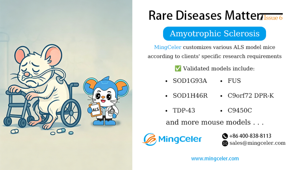
Amyotrophic Lateral Sclerosis (ALS), commonly known as Lou Gehrig’s disease, is a fatal neurodegenerative disorder characterized by motor neuron degeneration. Its clinical manifestations include progressive muscle atrophy, fasciculations, bulbar palsy, and pyramidal tract damage, ultimately leading to respiratory failure and death.
ALS is classified into sporadic ALS (SALS, 90%–95% of cases) and familial ALS (FALS, 5%–10% of cases), mostly inherited in an autosomal dominant pattern. The global prevalence of ALS is approximately 5 per 100,000 people, with a lifetime risk of 1 in 400–800. The peak age of onset is 55–75 years, and the median survival is about 27.5 months. In China, the average age of ALS onset is around 53.7 years, relatively younger than the global average.
Pathogenesis
The exact pathogenesis of ALS remains unclear, with several key mechanisms implicated:

1. Gene Mutation
- In Europe, mutations in the C9orf72 gene are the most common cause of familial ALS, accounting for 30%–40% of cases. These mutations exert neurotoxic effects via abnormal RNA aggregates and dipeptide repeat proteins (DPRs).

- In Asia, SOD1 gene mutations are the most prevalent, responsible for 20% of familial ALS and 2% of sporadic ALS. Mutations in FUS, C9orf72, and TARDBP are also common. SOD1 mutations cause structural abnormalities in superoxide dismutase (SOD1), forming misfolded aggregates that trigger oxidative stress and mitochondrial dysfunction.
- The TARDBP gene encodes TAR DNA-binding protein 43 (TDP-43), which maintains mRNA stability, protein translation, and nucleocytoplasmic transport. Loss or overexpression of TDP-43 protein disrupts motor neuron function.
2. Protein Homeostasis Imbalance
ALS-related pathogenic gene variants lead to protein misfolding, abnormal aggregation, or impaired degradation. These abnormal protein aggregates accumulate, exert neurotoxicity, and ultimately cause motor neuron degeneration and death.
3. Glutamate Excitotoxicity
Glutamate is the major excitatory neurotransmitter in the central nervous system (CNS). Synthesized at presynaptic terminals and diffusing across the synaptic cleft, it activates postsynaptic receptors and triggers action potentials. Overactivation or prolonged activation of glutamate receptors causes neuronal degeneration and death.
4. Oxidative Stress
SOD1 gene mutations alter antioxidant enzyme activity, disrupting the balance between intracellular oxidation and antioxidant systems. Free radicals attack neuronal cell membranes, proteins, and DNA, resulting in cellular damage.
Additionally, mitochondrial dysfunction, axonal transport abnormalities, neuroinflammation, and neurotrophic factor deficiency also contribute to ALS pathogenesis.
Gene Therapy
1. Antisense Oligonucleotide (ASO) Therapy
ASOs are short synthetic DNA molecules that bind to specific mRNAs and reduce target mRNA levels via RNase H-mediated degradation. In ALS therapy, ASOs are designed to target mutant SOD1 mRNA, reducing toxic protein production. For example, Tofersen is approved for treating SOD1-mutant ALS patients, reducing toxic protein via intrathecal injection. Jacifusen (targeting FUS mutations) has also shown efficacy in clinical trials.

2. AAV Gene Therapy
- SNUG01 (targeting the TRIM72 gene), delivered via AAV9 vectors, exerts pleiotropic effects including membrane repair and antioxidant activity, covering sporadic ALS. It has obtained FDA orphan drug designation and clinical trial approval.
- KLTO-202 (targeting soluble KL protein) alleviates inflammation and oxidative stress via AAV-mediated gene delivery, prolonging survival in ALS models, and has obtained FDA orphan drug designation.
3. Gene Editing Therapy
Gene editing technology can precisely excise the mutant SOD1 gene. In vitro experiments and animal model studies confirm its efficacy in reducing mutant SOD1 expression with minimal impact on wild-type SOD1, demonstrating therapeutic potential.
ALS Mouse Models
1. SOD1G93A Mice
Carry a human SOD1 gene with the G93A point mutation, mimicking human SOD1-mutant ALS. Onset occurs at 3–4 months of age, presenting with hindlimb weakness, tremors, and weight loss. Disease progression is rapid after onset, with an average survival of 120–130 days.
2. SOD1H46R/H48Q Mice
Exhibit progressive motor dysfunction, including weight loss, muscle weakness, and paralysis, with an average lifespan of approximately 189 days or shorter.
3. TDP-43 Mice
Display motor dysfunction, neuronal loss, and abnormal TDP-43 protein aggregation, recapitulating key pathological features of human ALS.
4. FUS Mice
Exhibit rapid disease progression with prominent RNA metabolism abnormalities.
5. C9450C Mice
Express 450 repeat sequences, used to study the relationship between autophagy defects and DPR accumulation.
6. C9orf72 DPR-KI Mice
Characterized by motor neuron loss, cortical hyperexcitability, behavioral deficits (e.g., anxiety-like behavior), and reduced C9orf72 protein expression.
MingCeler Biotech Supports Gene Therapy Development
Gene therapy offers hope for rare diseases, but its development and validation rely heavily on animal models. Leveraging its proprietary TurboMice™ Technology, MingCeler Biotech has developed multiple rare disease mouse models.
TurboMice™ Technology overcomes the limitations of long modeling cycles and low success rates for complex models. It enables editing at nearly any target gene locus and generates complete homozygous gene-edited mouse models directly from embryonic stem cells in as little as 2 months.
MingCeler Biotech customizes a full spectrum of ALS mouse models per client requirements, including SOD1G93A, SOD1H46R/H48Q, TDP-43, FUS, C9450C, and C9orf72 DPR-KI mice. We welcome researchers to contact us for inquiries!