




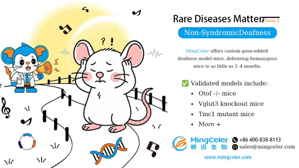
Deafness is one of the most common congenital birth defects. Worldwide, the incidence of hearing loss in newborns is 1.86‰, and more than 60% of deafness cases are attributed to genetic factors. Hereditary deafness is classified into two categories: Syndromic Hearing Loss (SHL) and Non‑Syndromic Hearing Loss (NSHL). NSHL is the most prevalent form, accounting for approximately 70% of all hereditary deafness cases.
Non‑syndromic deafness mainly affects the function of the cochlea or auditory nerve without lesions in other tissues or organs. Clinically, it presents as sensorineural hearing loss, which may be present at birth or develop gradually during childhood or adulthood.
Pathogenesis and Gene Therapy
The pathogenesis of non‑syndromic deafness is complicated, involving mutations in multiple genes such as mtDNA 12S rRNA, SLC26A4, GJB2, GJB3, and GJB6. Inheritance patterns include autosomal recessive, autosomal dominant, X‑linked, and mitochondrial inheritance. Known genes associated with non‑syndromic deafness are grouped by function:
(1) Cytoskeletal Protein-Encoding Genes:
Actin and myosin are critical proteins that regulate the structure and function of stereocilia. Mutations in related genes such as ACTG1 and MYO7A can disrupt actin filament organization or impair myosin’s ability to hydrolyze ATP and slide along actin filaments, ultimately affecting ciliary motility.
(2) Cell Junction Protein-Encoding Genes:
Intercellular junctions in the inner ear are essential for maintaining ion and voltage balance between endolymph and perilymph. Mutations in gap junction genes like GJB2 and GJB6 cause impaired potassium recycling, reducing or eliminating the endocochlear potential and leading to hair cell death.
Studies report that GJB2 mutations account for 10%–25% of non-syndromic hearing loss. Connexin 26, encoded by GJB2, is expressed in cochlear supporting cells and mediates intercellular communication. Common GJB2 mutations in the Chinese population include: c.235delC, c.299-300delAT, c.176-191del16, and c.109G>A.
(3) Ion Channel Protein-Encoding Genes:
Hearing loss-associated ion channel genes include SLC26A4 and TMC1, which play key roles in maintaining the ionic and electrical stability of inner ear fluids.
(4) Extracellular Matrix Protein-Encoding Genes:
The TECTA gene encodes tectorin alpha, a major structural component of the tectorial membrane.
(5) Hair Cell Synaptic Function Proteins:
Mutations in OTOF disrupt the function of hair cell synaptic proteins, resulting in auditory neuropathy. Otoferlin, encoded by OTOF, is a calcium-binding protein specific to cochlear ribbon synapses. Acting as a calcium sensor, it drives vesicle exocytosis, membrane fusion, and replenishment of synaptic vesicles at the active zone. OTOF mutations cause autosomal recessive deafness DFNB9, typically presenting as prelingual, moderate-to-severe hearing loss, often with temperature sensitivity.
The SLC17A8 gene encodes VGLUT3, which mediates glutamate uptake into synaptic vesicles in inner hair cells. It is critical for auditory pathway development and signal encoding. Mutations reduce synaptic glutamate levels, impairing action potential generation and auditory signal transmission, leading to autosomal dominant non-syndromic deafness DFNA25, characterized by progressive high-frequency hearing loss.
(6) Transcription Factor-Encoding Genes:
Genes such as POU3F4 and POU4F3 encode POU domain transcription factors that play essential roles in inner ear development. Additionally, many genes related to ciliary function and cellular homeostasis are involved, as well as genes encoding inner ear-specific RNAs, including 12S rRNA (encoded by mitochondrial DNA) and miRNA-96 (encoded by MIR96).

Gene Therapy for Hereditary Deafness
Rapid advances in gene therapy have brought new hope for rare diseases by targeting the genetic root causes. Three core strategies are used: gene replacement, gene suppression, and gene editing.
In preclinical research, more than 40 studies have successfully restored hearing in animal models for over 20 deafness‑causing genes. Three clinical trials for hereditary deafness have been approved globally, including the world’s first in vivo dosing for OTOF‑related deafness led by Prof. Shu Yilai at Eye & ENT Hospital of Fudan University. Gene therapy is poised to become a standard treatment for non‑syndromic deafness.

MingCeler Empowers Gene Therapy R&D
Gene therapy relies on validated animal models. Using proprietary TurboMice™ Technology, MingCeler has developed multiple rare disease mouse models. This ultra‑rapid generation of mouse models overcomes long timelines and low success rates of conventional methods, enabling in situ precision gene editing at nearly any locus.
MingCeler delivers homozygous mice directly from embryonic stem cells (ESCs) in as little as 2 months, with no allelic segregation distress and high genetic integrity.
We provide custom gene‑edited deafness mouse models, including:
- Otof⁻/⁻ mice
- Vglut3 knockout (KO) mice
- Tmc1 mutant mice
Reference
[1] Bu Y, Lü X, Wang Y, et al. Analysis of pathogenic gene mutations in 22 patients with non-syndromic hearing loss. Journal of Nanjing Medical University. 2010;30(3):390-393.
[2] Jiang L, Wang D, He Y, Shu Y. Advances in gene therapy hold promise for treating hereditary hearing loss. Molecular Therapy. 2023;31(4):934-950. doi:10.1016/j.ymthe.2023.02.001. PMID: 36755494; PMCID: PMC10124073.
[3] Sun Y, Jin C, Feng B, et al. Current status of gene therapy for auditory neuropathy. Chinese Journal of Otorhinolaryngology Head and Neck Surgery. 2024;59(5):510-518. doi:10.3760/cma.j.cn115330-20231029-00177.
[4] Chinese Journal of Medical Genetics. 2020;37(3):269-276. doi:10.3760/cma.j.issn.1003-9406.2020.03.008.
Advantages of MingCeler Custom Animal Model Services
1. Ultra-fast Timeline
MingCeler can generate custom mouse models in as short as 2 months. All founder mice are 100% homozygous, with no need for subsequent breeding and screening.
2. Higher Precision
Leveraging TurboMice™ technology, we perform in-situ precise gene editing at the embryonic stem cell level with strong tissue specificity. The established mouse models perfectly meet the demands of novel drug research and development.
3. Higher Efficiency
F0 generation mice generated via TurboMice™ are ready-to-use target models. All mice in the same batch derive from single cells with excellent genetic consistency. No chimeric mice are produced, delivering significantly higher efficiency.
4. Diversified Options
Traditional techniques are limited in mouse strain selection, while TurboMice™ offers far greater flexibility. A wide range of inbred and outbred strains are available, including Balb/c, ICR, C57BL/6, etc.
Powered by MingCeler’s proprietary EnhancerPlus technology platform and high-efficiency tetraploid complementation platform, we support flexible selection of gene editing loci, fragment lengths and editing numbers, free from allelic segregation issues.
Rapid Custom Mouse Models
- TurboMice™ Rapid Humanized Homozygous Mouse Customization
- TurboMice™ Rapid KO Homozygous Mouse Customization
- TurboMice™ Rapid Point Mutation Mouse Customization
- TurboMice™ Rapid KI Homozygous Mouse Customization
- TurboMice™ Rapid CKO Homozygous Mouse Customization
- TurboMice™ Rapid Homozygous Mouse Customization
Complex Custom Mouse Models
- TurboMice™ Multi-locus Gene Editing Mouse Customization
- TurboMice™ Long-fragment Gene Editing Mouse Customization
Ready-to-Use QuickMice™ Models
- QuickMice™ LaminA Progeria Mice
- QuickMice™ nuNPG Immunodeficient Mice
- QuickMice™ HPV Model Mice
- QuickMice™ HLA Humanized Mice
- QuickMice™ PCSK9 Humanized Mice
- QuickMice™ TIGIT Humanized Mice
- QuickMice™ ACE2 Model Mice